General Information about Tadora
Tadora is a drugs that belongs to a category of medicine known as phosphodiesterase-5(PDE5) inhibitors. It works by bettering blood move to the penis, permitting males to achieve and keep an erection. Tadora is simply prescribed for men over the age of 18 and is used to handle all forms of ED.
It is crucial to consult a doctor before taking Tadora to make sure it's safe for you. Your physician will consider your medical history and another medicines you take to determine if Tadora is the proper choice for you. Some folks might experience unwanted effects corresponding to headaches, dizziness, and upset abdomen while taking Tadora. It is important not to take Tadora with certain drugs, particularly these containing nitrates, as this can trigger a harmful drop in blood strain.
It is essential to observe your physician's instructions when taking Tadora. Do not take greater than the prescribed dosage, and don't take it more than once a day. If you expertise any discomfort or extreme unwanted aspect effects whereas taking Tadora, it is very important search medical attention instantly.
In conclusion, Tadora is a medication prescribed for managing all types of erectile dysfunction in men over 18 years old. It works by bettering blood circulate to the penis and can provide men with a firmer and longer-lasting erection. While it is an efficient treatment for ED, it is important to seek the assistance of a physician earlier than taking Tadora and to comply with their directions fastidiously. With the assistance of Tadora, males can confidently take pleasure in a satisfying sex life.
Tadora must be taken 30 minutes to an hour before sexual activity. The effects can last as long as 4 hours, giving men a adequate window of time to engage in sexual activity. However, Tadora will solely work when a person is sexually aroused, so it is important to observe that it's not an aphrodisiac.
Tadora has been proven to be an efficient and protected therapy for ED. In fact, in a scientific research, 81% of males who took Tadora reported improved erections. It is necessary to note that Tadora does not treatment ED, but it could possibly effectively manage it. This signifies that the medicine must be taken every time a person needs to have interaction in sexual exercise.
One of the principle causes of ED is a decreased blood flow to the penis. This could be caused by quite lots of factors similar to diabetes, high blood pressure, and smoking. Tadora helps to loosen up the blood vessels within the penis, which permits for increased blood move and ultimately, a firmer and longer-lasting erection.
Erectile dysfunction (ED) is a typical condition that affects tens of millions of men worldwide. It is the lack to achieve or preserve an erection sufficient for sexual exercise. Although it could be a difficult and embarrassing matter to discuss, ED is a treatable situation. One medicine that is commonly prescribed for managing ED is Tadora.
Details the surveillance of influenza symptoms impotence of organic origin tadora 20 mg with amex, vaccination options, and possible complications. Provides a comprehensive review of disease and medication management for major body systems, including management of the common cold and flu. Infectious Diseases and Conditions Web Sites of Interest National Institute of Allergy and Infectious Diseases · 733 1887, and a young Marine Hospital Service physician named Joseph Kinyoun. As Kinyoun and others began to realize, immigrants arriving on the shores of the United States often brought with them infectious diseases, such as cholera and yellow fever. Kinyoun, who had seen science research centers in Europe, had also learned about the new science of bacteriology and put its principles to use by screening newly arrived immigrants at the bacteriological laboratory he founded on Staten Island. The Edge of Discovery: A Portrait of the National Institute of Allergy and Infectious Diseases. National Research Council, Committee on the Organizational Structure of the National Institutes of Health. Rather than simply depend on symptoms for diagnostic purposes, Kinyoun discovered that culturing cholera bacteria was a reliable way to determine if immigrants to the United States had cholera. The name of the lab became the National Institute of Health in 1930, the same year that fellowships were started there. In 1948, the agency became known as the National Institutes (plural) of Health, and specialty areas were split into institutes, which have increased in number since and which are now located in Bethesda, Maryland. Other research has found a vulnerable area on the virus, a finding that could lead to the development of a vaccine. A major initiative for development of drug treatments for tuberculosis is ongoing, leading to the identification of some promising drugs to treat the disease. Genome sequences of viruses and bacteria, such as Plasmodium, which causes malaria, are currently being utilized to develop better vaccines. Collaborations are forged between established researchers in the United States and scientists in other Centers and Institutes of the National Institutes of Health Center for Information Technology Center for Scientific Review Eunice Kennedy Shriver National Institute of Child Health and Human Development John E. Necrotizing fasciitis Category: Diseases and conditions Anatomy or system affected: Skin, tissue Also known as: Flesh-eating bacteria, streptococcal gangrene Definition Necrotizing fasciitis is a rapidly progressive and aggressive rare infection of fascia and soft tissue that can follow minor trauma or surgery or may occur without any known cause. The term "necrotizing fasciitis" was first used in 1952 to describe a quickly spreading soft tissue infection. The disease has likely existed for centuries and was well documented during the American Civil War. Although often caused by group A beta-hemolytic streptococci bacteria, the disease, which leads to decaying (gangrenous) skin, can also be caused by many other bacteria and is usually caused by mixed bacterial infection. Prompt diagnosis guided by a high index of suspicion is the key to successful treatment. Causes Group A streptococcal infection also causes impetigo and strep throat, which are less serious than Infectious Diseases and Conditions necrotizing fasciitis. M-protein serotypes of these bacteria may be responsible for the more aggressive necrotizing fasciitis. In most cases, necrotizing fasciitis is caused by a mixed bacterial infection involving aerobic and anaerobic species. Common identified bacterial species include methicillin-resistant Staphylococcus aureus, Escherichia coli, Pseudomonas, Clostridium, Klebsiella, Proteus, Vibrio, and Bacteroides. Entry into the body may occur from trauma as minor as a scratch, insect bite, burn, or needle puncture. Once in the subcutaneous tissue, the infection spreads along facial plains and move deeper into soft tissues to involve muscle and fat. Enzymes and toxins produced by the bacteria may cause vascular occlusion, resulting in a loss of oxygen, tissue necrosis, and toxic shock. Risk Factors Any person at any age can be affected by necrotizing fasciitis, but persons with compromised immune systems or with certain underlying conditions are at higher risk. These risk factors include intravenousdrug use, alcoholism, human immunodeficiency virus infection, diabetes, peripheral vascular disease, cancer, renal failure, liver disease, and treatment with chemotherapy or corticosteroids. Symptoms Early symptoms of necrotizing fasciitis include an unusual amount of pain in an area of a recent injury. Symptoms of generalized illness, such as fever, nausea, and weakness, may soon follow. A cracking noise or sensation beneath the skin (crepitus) may Necrotizing fasciitis · 737 be present. Signs of toxic shock may include a rapid drop in blood pressure and loss of consciousness. Although laboratory studies, tissue cultures, biopsy results, and imaging studies may aid in the diagnosis of necrotizing fasciitis, a presumptive diagnosis may need to be made on history and physical examination alone. The absolute necessity of prompt treatment precludes the need to wait for results of supporting diagnostic tests. Early signs and symptoms can be deceptive, so a high index of suspicion is the key to diagnosis. Laboratory testing may include a complete blood count that shows elevated white blood cells. Helpful imaging studies include a computed tomography scan, magnetic resonance imaging, and an ultrasound. Treatment and Therapy Necrotizing fasciitis is a medical emergency and may require treatment in an intensive care setting. Intravenous fluid administration, medication to control blood pressure, oxygen, cardiac monitoring, and intubation may all be necessary in a person presenting with toxic shock. Broad-spectrum antibiotics should be started without waiting for the results of blood or tissue cultures. Some commonly used antibiotics for this infection are penicillin, clindamycin, vancomycin, and cephalosporins.
Higher risk is associated also with old age erectile dysfunction jelly tadora 20 mg order visa, immunosuppressive therapy or glucocorticoid therapy, and intravenous drug abuse. Symptoms the symptoms of a mycotic aneurysm vary according to their size and their site. If the site of the mycotic aneurysm is bleeding, there will be additional symptoms. The symptoms include fever; abdominal, thigh, neck, or arm pain; palpable mass; nausea; weakness; and fatigue. If the mycotic aneurysm is in the arteries of the brain, the symptoms will be headache, seizures, bleeding into the brain, and nausea and vomiting. Infectious Diseases and Conditions Screening and Diagnosis There is no routine screening for a mycotic aneurysm. The diagnosis is based on the symptoms, on increased white blood cells, and on diagnostic imaging. Treatment and Therapy the treatment for mycotic aneurysm is antibiotics and surgery. If the bacterium has not been identified, antibiotics will be chosen based on the likely bacteria. A brain mycotic aneurysm sometimes requires that the aneurysm be blocked off with tiny metal coils to prevent rupture. Mycotic aneurysms require surgery to remove the infective debris and to replace or bypass the damaged artery. It is thought that avoiding Salmonella infections of the gastrointestinal tract can decrease the likelihood of contracting a Salmonella infection, including a mycotic aneurysm, in other parts of the body. Salmonella infections are contracted by contact with infected chickens, pigs, and eggs. Causes Many cases of myocarditis have no identifiable cause and are called "idiopathic myocarditis. Toxic myocarditis is caused by drugs such as chemotherapeutic drugs, lithium, or cocaine; by heavy metals such as copper, iron, or lead; by toxic substances such as arsenic, carbon monoxide, or other inhalants; and by physical agents such as electric shock or radiation. Treatment and Therapy the universally recommended therapy for myocarditis is bed rest, no physical activity, and supplemental oxygen. Corticosteroids may be given to help inflammation, and the patient will most likely be admitted to a hospital. Forinstance,ifthecauseisabacterial infection, the doctor will prescribe antibiotics; if the cause is viral, the doctor will prescribe antiviral agents. Immunosuppressive therapy may be used if the myocarditis is caused by an autoimmune disorder such as lupus or scleroderma. If heart failure symptoms are present, the doctor will prescribe medications to support the function of the heart. To help reduce the chance of getting myocarditis, one should reduce exposure to identified causes. The following symptoms may appear slowly or suddenly: flulike complaints, including fever, fatigue, muscle pain, vomiting, diarrhea, and weakness; a rapid heart rate; chest pain; shortness of breath and respiratory distress; and a loss of consciousness. To do this, a doctor will ask the patient about symptoms and medical history and will perform a physical exam. Risk Factors Generally, women are affected more often than men, although inclusion-body myositis affects twice as many men as women. Polymyositis is observed in persons between twenty and sixty years of age, whereas inclusion-body myositis is more common after age fifty years. African Americans are at higher risk for myositis, while the lowest rates of myositis are reported in persons of Japanese origin. Symptoms Common symptoms of the inflammatory myopathies include muscle weakness, sometimes with muscle pain, that lasts for more than a few weeks; general tiredness and fatigue; difficulty climbing stairs, standing up from a seated position, or reaching up; and difficulty swallowing. Additional symptoms for the various myopathies include a variety of skin symptoms (such as a rash or scaly, dry, and rough skin) in dermatomyositis; and hardened lumps of calcium (calcinosis) under the skin in juvenile dermatomyositis. Unlike other inflammatory myopathies, the muscle weakness in inclusion-body myositis is often asymmetrical. Treatment and Therapy Treatment for myositis generally includes rest, physical therapy, and the use of anti-inflammatories (corticosteroids as first-line therapy and methotrexate, hydroxychloroquine, and azathioprine), and intravenous immunoglobulin. Myositis Category: Diseases and conditions Anatomy or system affected: Muscles, musculoskeletal system Also known as: Idiopathic inflammatory myopathy, inflammatory myopathy Definition Myositis is a general term for a group of rare chronic conditions characterized by inflammation of the skeletal muscles. Myositis refers to the inflammatory myopathies, including polymyositis, dermatomyositis, inclusion-body myositis, and juvenile myositis. Inflammatory myopathies can also be caused by certain medications or by exposure to a toxic substance; these myopathies are usually not chronic and resolve once the harmful substance is removed. It is believed that an environmental factor, such as a viral infection, triggers myositis in people who might be genetically predisposed to the condition. The damage in myositis is 730 · Myositis Prevention and Outcomes Because the cause of myositis is unknown, there is no known way to prevent the condition. To lessen the severity of dermatomyositis, however, persons with the condition should avoid excessive exposure to the sun, which can worsen any dermatomyositis-associated skin rashes. Myositis: A Medical Dictionary, Bibliography, and Annotated Research Guide to Internet References. Bordetella pertussis and Corynebacterium diphtheriae, sometimes found in the nasopharynx, cause severe upper respiratory infections but are infrequent in the United States because of immunizations.
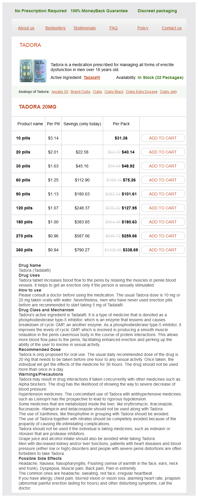
Tadora Dosage and Price
Tadora 20mg
- 10 pills - $31.36
- 20 pills - $40.14
- 30 pills - $48.92
- 60 pills - $75.26
- 90 pills - $101.61
- 120 pills - $127.95
- 180 pills - $180.63
- 270 pills - $259.66
- 360 pills - $338.69
This test is nearly as accurate as a blood test erectile dysfunction herbal remedies 20 mg tadora order with amex, and because it does not involve a needle stick, it is favored by many persons. Nonnucleoside reverse transcriptase inhibitors inhibit the virility of retroviruses and cause cell death. Prevention and Outcomes the best way to prevent infection by a retrovirus is to avoid contact with that virus. The author provides a detailed account of virus structure and replication and of the basis for disease pathology. A minimum of nine endogenous retroviruses are present in vertebrate genomes, accounting for approximately 1 percent of the human genome, and they appear latent, with no effect on the hosts. Exogenous retroviruses, which are passed among vertebrates through blood and bodily secretions, can be more pathogenic. All seven genera of the two subfamilies of retroviruses share the same mode of replication and basic virion structure, and they spread through host cells rapidly. Retroviruses consist of a lipid envelope that surrounds a spherical and electrondense protein core, or capsid. Immature viral particles are then released from the infected host cell to spread throughout the body. Retroviruses can evolve rapidly and repeatedly on the basis of selective needs, resulting from immune attacks or administered drugs. Retroviridae · 919 Pathogenicity and Clinical Significance Outcomes of pathogenic retroviral infections are host specific and may be neurologic, immunodeficient, or wasting. Because retroviruses can change the host genome, they also can develop oncogenes in the body. The particular drugs used vary around 920 · Reverse transcriptase inhibitors a standard, recommended regimen; medications are added, removed, or replaced as necessary when resistance builds to a certain drug or entire mechanistic class. Because retroviral genomes have the ability to change and adapt to outside pressures rapidly and efficiently, drug susceptibility varies by viral strain and by particular infected host, and it depends on immune system attacks and on treatments administered to the host. If, after twenty days on a regimen, viral load is not lowered to undetectable levels, the regimen is considered ineffective and can lead to virologic failure without a change of therapy. A thorough discussion of retroviral families and pathogenicity, with descriptions of the resultant diseases. A textbook discussion on retrovirus transmission that is particularly focused on transmission among children. To do this, it Infectious Diseases and Conditions uses an enzyme called reverse transcriptase. Treatment and Therapy the goals of treatment are to kill the strep bacteria, treat the inflammation caused by the rheumatic fever, and prevent future cases of rheumatic fever. Treatment includes medication to treat the strep infection (such as penicillin or other antibiotics, including erythromycin and azithromycin). The patient may have to be on bed rest or restricted activity for a period of time. Prevention and Outcomes One should treat strep throat with antibiotics promptly to help prevent the onset of rheumatic fever. Persons with a sore throat and fever that last more than twenty-four hours should consult a doctor. Rheumatic Fever in America and Britain: A Biological, Epidemiological, and Medical History. Rheumatic fever Category: Diseases and conditions Anatomy or system affected: All Definition Rheumatic fever is an inflammatory condition. The most severe complication is rheumatic heart disease, which may permanently damage the heart valves, which affect the flow of blood to and from the heart. Often, the symptoms of valve damage appear ten to thirty years after the initial event. Causes Rheumatic fever is caused by the immune system responding to group A Streptococcus pharyngitis (strep throat). In this case, the immune system not only fights the bacteria but also attacks its own tissue, often heart tissue. Risk Factors Factors that may increase the risk of rheumatic fever include previously having rheumatic fever, being malnourished, and living in overcrowded conditions. Symptoms Symptoms usually appear two to four weeks after a strep infection and include pain and swelling in large joints; fever; weakness; muscle aches; shortness of breath; chest pain; nausea and vomiting; hacking cough; circular rash; lumps under the skin; and abnormal, sudden movements of arms and legs. Screening and Diagnosis A doctor will ask about symptoms and medical history and will perform a physical exam that includes a careful examination of the heart. The doctor may take a throat culture and order a blood test to check for streptococcal antibodies. Rhinosporidiosis · 923 trachea (windpipe), bronchi, ears, scalp, genitals, rectum, and the skin. Causes Rhinosporidiosis is caused by the organism Rhinosporidium seeberi, which has features of both fungi and protozoa. Risk Factors the greatest number of rhinosporidiosis cases has been reported in India and Sri Lanka, but the infection has been diagnosed in persons from the Americas, Europe, Africa, and Asia. The reservoir of this microorganism and the mode of transmission are unknown, although R. These tumors can obstruct the nose and cause increased nasal drainage, cough, sneezing, and postnasal discharge. The tumors in the eye can cause excessive tearing, redness, photophobia, and infection. Screening and Diagnosis There is no routine screening for rhinosporidiosis, but because of the external location of the growths, they are easy to see.
© 2025 Adrive Pharma, All Rights Reserved..