General Information about Super P-Force Oral Jelly
In conclusion, Super P-Force Oral Jelly is a game-changer on the planet of male sexual well being, offering a convenient and effective answer for males with each ED and PE. Its quick motion, pleasant taste, and dual-action make it a well-liked selection among men seeking to improve their sexual performance. However, it could be very important use it responsibly and solely as directed by a healthcare skilled to make sure its safety and effectiveness.
On the opposite hand, Dapoxetine Hydrochloride is a relatively new treatment used to deal with untimely ejaculation. It belongs to the category of selective serotonin reuptake inhibitors (SSRIs) which work by rising the degrees of serotonin within the brain. Serotonin is a neurotransmitter that plays a crucial position in controlling ejaculation and by increasing its levels, Dapoxetine helps in delaying ejaculation, permitting men to have higher management over their sexual exercise.
Another good thing about Super P-Force Oral Jelly is its nice taste. The jelly comes in numerous fruit flavors such as strawberry, orange, mango, and banana, making it a extra palatable choice for these who are not keen on the bitter style of conventional erectile dysfunction medication.
Super P-Force Oral Jelly works by combining the consequences of these two energetic elements, making it a strong and effective therapy for both ED and PE. It is a handy and cost-effective answer for men who're affected by each situations as they will now take just one medicine as a substitute of two.
The primary energetic elements in Super P-Force Oral Jelly are Sildenafil Citrate and Dapoxetine Hydrochloride. Sildenafil Citrate, also called Viagra, is a well-known treatment used to deal with ED. It works by rising blood circulate to the penis, resulting in an extended lasting and firmer erection.
Super P-Force Oral Jelly is a protected and effective remedy for ED and PE, with minimal unwanted aspect effects similar to headache, dizziness, and flushing. However, some males might experience extra extreme unwanted effects such as blurred vision, adjustments in listening to, and priapism (painful erection lasting longer than 4 hours). If any of these occur, you will need to seek medical attention immediately.
One of the primary benefits of Super P-Force Oral Jelly is its quick action. Unlike traditional tablets, the jelly form of this medicine is quickly absorbed by the body, allowing the energetic ingredients to take effect within 15-20 minutes. This makes it a perfect therapy for males who wish to be spontaneous in their sexual activities.
As with any treatment, there are some precautions that must be taken when using Super P-Force Oral Jelly. It isn't recommended for men with a historical past of coronary heart, kidney, or liver problems. It should also not be taken with different medications that include nitrates as it could trigger a sudden drop in blood pressure. It is all the time finest to consult with a doctor before taking any new medicine, especially in case you have any underlying well being issues.
Super P-Force Oral Jelly is a revolutionary medication that has been specially designed to tackle two of the commonest male sexual health problems - premature ejaculation and erectile dysfunction. Its unique jelly kind makes it easier to swallow and has gained recognition among men who've problem taking tablets or tablets.
Most episodes of contrast-induced nephrotoxicity are mild and characterized by a reversible 1- to 3-mg/dL rise in serum creatinine; dialysis therapy is rarely needed and usually only in patients whose baseline serum creatinine level is high drugs for erectile dysfunction in nigeria discount super p-force oral jelly 160 mg line, for example, > 3 mg/dL. In terms of being an absolute measure, serum creatinine is an unreliable measure of renal function. Alternative if an emergency procedure is required: 5-mL/kg bolus of normal saline 1 hr before and 1 mL/kg per hr for 12 hr after the procedure. Alternative fluid regimen with bicarbonate: add 154 mL of 1000 mEq/L sodium bicarbonate to 850 mL of 5% dextrose in water (D5W) (or add 3 ampules of standard bicarbonate to 1 L D5W). Initial bolus of 3 mL/kg for 1 hr before injection of contrast material, followed by 1 mL/kg per hr for 6 hr after the procedure. An exception involves the pre-6000 series StarrEdwards caged ball valves; devices rarely used now. The hazard primarily reflects the possibility of deflecting the foreign body sufficiently to injure vital structures. Dental alloys, wires, splints, dental braces, and prostheses do not appear to pose a risk to the patient, although such material may result in artifactual changes. Cutaneous burns can result from contact of the skin with metal objects, including neurosurgical halo pins, pulse oximetry probes, and drug-eluting medical patches that contain metal foil. Many bullets are safe, but those with metal (specialized bullets, such as metal jackets) may pose a risk. Bentur Y, Horlatsch N, Kiren G: Exposure to ionizing radiation during pregnancy: perception of teratogenic risk and outcome. International Commission on Radiological Protection: Pregnancy and medical radiation. Fattibene P, Mazzei F, Nuccetelli C, et al: Prenatal exposure to ionizing radiation: sources, effects and regulatory aspects. Dunn K, Yoshimaru H, Otake M, et al: Prenatal exposure to ionizing radiation and subsequent development of seizures. In Occupation and environmental reproductive hazards: a guide for clinicians, Baltimore, 1993, Williams & Wilkins, p 165. Giles D, Hewitt D, Stewart A, et al: Malignant disease in childhood and diagnostic irradiation in utero. Wakeford R: Childhood leukemia following medical diagnostic exposure to ionizing radiation in utero or after birth. Bona G, Zaffaroni M, Defilippi C, et al: Effects of iopamidol on neonatal thyroid function. Mallick S, Petkova D: Investigating suspected pulmonary embolism during pregnancy. Nikolaou K, Thieme S, Sommer W, et al: Diagnosing pulmonary embolism: new computed tomography applications. Perrier A, Desmarais A, Goehring C, et al: d-Dimer testing for suspected pulmonary embolism in outpatients. Regardless of the procedure, some anticoagulated patients are at potential significant risk of hemorrhage from the procedure. However, emergency reversal of anticoagulation in order to perform the procedure may also place the patient at risk for serious thrombotic complications. If the procedure is not needed for life-saving therapy, postponing the procedure or providing empiric treatment may be a reasonable choice. In contrast, emergency procedures to reverse an imminent life-threatening condition should never be withheld and emergency reversal of anticoagulation may be required. Although laboratory tests to determine drug presence, drug concentration, and level of anticoagulant effect can be useful in the assessment of bleeding risk, standard coagulation assays accurately monitor the degree of anticoagulation for only a few agents. The direct measurement of drug concentration is not suitable in clinical practice because of the time required to perform the laboratory analysis. Normal thrombin time is suitable for excluding significant dabigatran levels but too sensitive for determining the degree of anticoagulant effect. Unfortunately, there are no validated systems available to quantify risk of bleeding. These patients include those with a recent diagnosis of pulmonary embolism, significant clot burden, or those with mechanical hardware such as a prosthetic cardiac valve. When clinically feasible, it is best to allow the anticoagulant effect of a medication to wane rather than emergently reversing the medication in these patients. However, life-saving measures such as thoracostomy, central venous catheterization, and endotracheal intubation are sometimes required even in these severe settings. Additional life-saving procedures that may carry a high risk of bleeding in the setting of anticoagulation, but should not be withheld, include defibrillation and pericardiocentesis. The gravity of the clinical scenario is important to keep in mind when weighing the risk of performing a procedure in the setting of anticoagulation. Furthermore, the simple act of controlling periprocedural bleeding may not entirely end the risk of serious harm. For instance, periprocedural bleeding with percutaneous coronary intervention is associated with an increased short- and long-term morbidity and mortality, including major adverse cardiovascular events and readmission rates well after the bleeding is controlled. Most importantly, never withhold emergency life-saving procedures such as endotracheal intubation, tube thoracostomy, cardiac defibrillation, pericardiocentesis, or vascular access when necessary. Stabilization with supportive treatments such as oxygenation, intravascular volume resuscitation, and repletion of blood products via transfusion are the initial steps of assessment and management of bleeding in the anticoagulated patient.
Hepatic Veins and Portal Venous System Hepatic veins Distribution of the hepatic veins Variations of the hepatic veins Collateral channels Portal vein Left gastric vein Right gastric vein Paraumbilical veins Cystic veins Splenic vein Short gastric veins Left gastroepiploic vein Pancreatic veins Inferior mesenteric vein Superior rectal veins Sigmoid veins Left colic vein Superior mesenteric vein Jejunal and ileal veins Ileocolic veins Right colic vein Middle colic vein Right gastroepiploic Pancreaticoduodenal veins Anastomoses between the portal and the systemic circulations Pancreatic venous system Hepatic Veins Distribution of the Hepatic Veins the hepatic veins drain the liver parenchyma and start as interlobular veins erectile dysfunction statistics worldwide super p-force oral jelly 160 mg purchase without prescription, draining the sinusoids of the hepatic lobules. According to the classical description, these end at the sublobular veins, which will drain into the hepatic veins There are three main hepatic veins emerging from the upper, posterior surface of the liver, opening at the inferior vena cava, and an individual vein from the caudate lobe. These are called the upper group, which consists of the large, right, middle, and left hepatic veins, and the smaller caudate lobe vein, delineating four well-defined territories of drainage. The right hepatic veins run at the right hepatic fissure, which divides the right hepatic lobe in the anterior and the posterior sectors. In 16 of 25 casts of liver specimens, there was only one right hepatic vein of large diameter receiving several tributaries from the several segments of the right lobe. There is a direct relationship between the right hepatic vein and the bifurcation of the main portal vein and the right branch of the portal vein. Outside the liver the portal vein crosses over the inferior vena cava, and the superior mesenteric vein follows a relatively parallel pathway along the inferior vena cava. One cast showed two right hepatic veins, parallel to each other and with similar sizes. In two cases, the right hepatic vein had a single short trunk of about 1 cm, but was bifid with two parallel veins peripherally. Six livers showed an accessory right hepatic vein, caudal and distal, but parallel to the main vein and posterior to the portal vein bifurcation. The distance from the anterior aspect of the right hepatic vein, at 1 cm from the inferior vena cava and the posterior aspect of the portal bifurcation, was measured in a straight line. The distance between the anterior aspect of the accessory right hepatic veins measured at 1 cm from the inferior vena cava and the portal bifurcation was 2. The middle hepatic vein joins the left hepatic vein to form a single venous trunk ending in the anterolateral aspect of the inferior vena cava. There was only one middle hepatic vein in all 25 cases of the series analyzed; this vein joined the inferior vena cava directly in about 20% of cases and formed a common trunk with the left hepatic vein in 80% of cases. The distance between the inferior aspect of the middle hepatic vein, at 1 cm from the inferior vena cava and the superior posterior aspect of the portal bifurcation and left portal trunk, was measured in a straight line. The caudate lobe vein is an independent tributary of the inferior vena cava and opens in the cava in a much lower position, in relation to the three main hepatic veins. The lower group of hepatic veins are smaller and numerous and drain liver parenchyma directly to the inferior vena cava, from the right liver lobe and the caudate lobe. Portal Vein the portal vein is about 7 to 8 cm in length and carries the visceral blood to the liver, where it ramifies following the segmental pattern, like the hepatic artery, reaching the sinusoids, from which the blood again converges to drain into the inferior vena cava through the hepatic veins. The portal vein results from the confluence of the splenic vein and the superior mesenteric vein, and follows a path, posteriorly to the pancreatic head and anteriorly to the inferior vena cava, reaching the liver through the lesser omentum and anterior to the epiploic foramen. Inside the lesser omentum and at the porta hepatis, it is posterior to the bile duct and the hepatic artery. The bile duct is parallel, but lateral, whereas the hepatic artery is parallel, but medial. The right portal branch enters the right hepatic lobe after receiving the cystic vein. At the left lobe of the liver, it is joined by the paraumbilical veins and the ligamentum teres (residuum of the obliterated left umbilical vein). There is also a connection with the inferior vena cava by the ligamentum venosum (remnant of the occluded ductus venosus). Variations of the Hepatic Veins There may be several large accessory hepatic veins draining the upper (diaphragmatic) part of the right and left liver lobes, joining the main hepatic veins, close to the outlet ring at the inferior vena cava. Several additional draining veins, from territories not usually drained by the three main veins, may be tributaries of the right, middle, and left hepatic veins. Accessory hepatic veins may also be encountered in the lower group, draining a variable amount of parenchyma from the right liver lobe, with an incidence of up to 15% of the population. Anatomy of the Portal Bifurcation the anatomy of the portal vein at the bifurcation is variable, and the bifurcation can be extrahepatic in about 25% of the cases. In 24 livers studied, the portal vein presented a very short and thin right trunk and a long left trunk. In almost every specimen, branches to the caudate lobe were seen arising from the bifurcation of the portal vein or from the first 1 cm of the left trunk. In another liver the hepatic artery bifurcated proximally and to the left of the portal bifurcation, giving off one large anterior branch and a smaller branch, which crossed posterior to the portal bifurcation and followed a path on the posterosuperior aspect of the right trunk for the portal vein. In one case there was a posterior arterial branch in the first 2 cm of the left portal trunk. In nine specimens there were arterial and/or biliary Venous Collateral Channels When obstruction of the hepatic veins or the suprahepatic segment of the inferior vena cava occurs, several collateral pathways develop and may be divided into three types. Extrahepatic Collaterals developed through the liver capsule toward retroperitoneal and intercostal veins. IntrahepaticInterlobar Collaterals developed from the hepatic segment with venous obstruction to adjacent patent hepatic veins. Indeterminate Type ("Spiderweb" appearance) Fine and/or coarse collateral networks emanating from the occluded vein (at phlebography), without certain flow direction. In these cases there was an intimate relationship between these two bilio-arterial structures, but overall the biliary branches were posterior. In general the biliary and arterial structures are close to the superior aspect of the portal trunks and more frequently are anterosuperior. Tributaries Besides the splenic and the superior mesenteric veins, other tributaries are the left gastric, right gastric, the paraumbilical, and cystic veins. A rare variation is the duplication of the portal vein, which may also be related to recanalized occlusion.
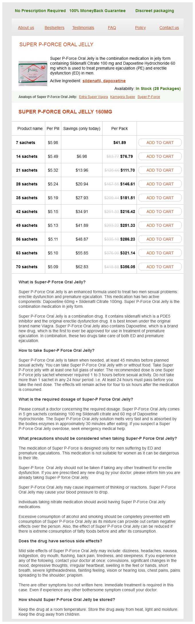
Super P-Force Oral Jelly Dosage and Price
Super P-Force Oral Jelly 160mg
- 7 sachets - $41.89
- 14 sachets - $76.79
- 21 sachets - $111.70
- 28 sachets - $146.61
- 35 sachets - $181.51
- 42 sachets - $216.42
- 49 sachets - $251.33
- 56 sachets - $286.23
- 63 sachets - $321.14
- 70 sachets - $356.05
Cold Gastric Lavage the stomach lies in close proximity to the liver erectile dysfunction karachi discount super p-force oral jelly 160 mg buy on line, great vessels, kidneys, and heart. The gastric mucosa is not subject to the intense vasoconstriction observed on exposure of the skin to ice water. Studies of cold gastric lavage in a canine model produced cooling rates five times greater than in controls exposed to ambient air at room temperature (0. A faster lavage rate can be maintained if suction is used to withdraw the instilled fluid. Connect this container directly to the lavage tubing and ideally allow passage of water but not ice, which may occlude the tube. Because large volumes of water are needed, it is helpful if additional ice can be added to the container without interrupting the lavage. A large syringe can be used as an alternative to gravity instillation, but this is usually slower. Use a standard lavage setup (for use in drug overdoses) and a largebore gastric tube. Using the clamps, intermittently instill ice water by gravity and withdraw it by suction. Complications A major potential complication of cold gastric lavage is pulmonary aspiration. Because of the large volume of water used and the frequent depression of airway reflexes seen with severe heatstroke, this technique should rarely be used in a patient who is not endotracheally intubated. If tap water is used, water intoxication, hyponatremia, and other electrolyte disturbances are potential complications, particularly in pediatric or geriatric patients. Water is absorbed from the stomach and, with large-volume lavage, may pass the pylorus into the small intestine. In canine studies, large-volume gastric lavage with tap water did not cause electrolyte abnormalities. Theoretically, passage of cold water through the esophagus, located directly behind the heart, has the potential to induce cardiac dysrhythmias. Dysrhythmias have not been observed in canine studies or in case reports of human heatstroke victims cooled with this technique. Peritoneal lavage is expected to exchange heat much faster than possible with gastric lavage. Peritoneal lavage achieves some of the fastest cooling rates ever reported in large animal or human studies (see Table 65. A case report of cooling via cold peritoneal lavage for hyperthermia after the ingestion of ecstasy demonstrated rapid cooling. Peritoneal lavage is used extensively to treat hyperthermia under various conditions and typically decreases core temperatures 5°C/hr to 10°C/hr (41°F/ hr to 50°F/hr). This cooling technique is relatively contraindicated by conditions that preclude placement of a lavage catheter. It can theoretically be combined with other techniques to speed cooling of heatstroke patients with refractory hyperthermia. As the most invasive cooling technique, it requires time, proper equipment, and surgical expertise to institute. Its use is probably best suited to situations in which heatstroke patients are not responding to external cooling and adequate equipment and personnel are readily available. Place a standard peritoneal lavage catheter (as for diagnostic use in trauma patients) via any of the techniques described in Chapter 43. Use of a larger peritoneal dialysis catheter may speed instillation and withdrawal of fluid. One approach is to instill and withdraw 500 to 1000 ml every 10 minutes until adequate cooling is achieved. Rectal temperature may be falsely low during lavage because of the presence of cold water around the rectum at the level of the rectal temperature probe. It may be preferable to monitor temperature in the tympanic membrane or esophagus when using this technique. Complications the potential complications of peritoneal lavage cooling are primarily related to placement of the catheter and include bowel or bladder perforation and placement into the rectus sheath rather than the peritoneum. Other Cooling Techniques "Rewarming" techniques are used to minimize ongoing heat loss via the respiratory tract in hypothermic patients. The use of dry, hot air to maximize evaporative heat loss from the lungs might cause respiratory complications. Hemodialysis or partial cardiopulmonary bypass could theoretically be used to cool heatstroke patients. A 2005 case report described successful treatment of a heatstroke patient with multiple-organ failure refractory to conventional cooling techniques with cold hemodialysis initially at 30°C (86°F) and later at 35°C (95°F), followed by continuous hemodiafiltration with cold dialysate (35°C [95°F]) at a high flow rate of 18,000 ml/hr. They circulate temperature-controlled sterile saline placed in the bladder or inferior vena cava. Although these devices have not been used in heatstroke patients, studies have found the cooling catheters to be very effective for neurologic conditions in both human and animal models. Cienki and colleagues demonstrated enhanced decreases in temperature with the administration of ketorolac, 30 mg intravenously. In the group receiving ketorolac, the average rectal temperature after 90 minutes was two times lower than in those receiving placebo saline (3. Conclusion Rapid cooling is the key step in the emergency management of heatstroke patients. Survival rates approach 90% when elevated temperatures are lowered in a timely fashion. It combines the advantages of simplicity and noninvasiveness with the most rapid cooling rates achieved with any external technique.
© 2025 Adrive Pharma, All Rights Reserved..