General Information about Prandin
In conclusion, Prandin is an efficient medication for managing sort 2 diabetes. By stimulating the pancreas to supply more insulin, it helps the body use glucose more successfully and retains blood sugar ranges in a wholesome range. Although it may trigger some unwanted effects, these can usually be managed with correct monitoring and changes. As at all times, it is essential to comply with a healthcare supplier's instructions and maintain a wholesome life-style while taking Prandin to achieve optimum leads to managing diabetes.
Like any treatment, Prandin might trigger unwanted effects in some people. The commonest unwanted aspect effects reported include low blood sugar (hypoglycemia), headache, dizziness, and gastrointestinal discomfort. These unwanted side effects are normally mild and can be managed by adjusting the dose or timing of the treatment. It is essential to observe blood sugar levels and report any side effects to a healthcare supplier.
One of the principle benefits of Prandin is its ability to control blood sugar levels shortly after a meal. This is particularly helpful for individuals who struggle with high blood sugar spikes after consuming. By taking Prandin before a meal, it may possibly forestall these spikes from occurring and help hold blood sugar ranges more steady throughout the day.
Prandin is prescribed for patients who have not been in a position to adequately control their blood sugar ranges by way of diet and train alone. It is usually taken before meals, as it really works by growing insulin production when glucose ranges rise after eating. By doing so, it helps the physique use glucose more successfully, maintaining blood sugar levels in a healthy range.
Another benefit of Prandin is that it has a shorter duration of action in comparison with different diabetes medications, which means its effects wear off faster. This may be helpful for individuals with irregular meal patterns or those that might skip meals, because it allows for more flexibility in when the treatment must be taken. However, it's still essential to hold up a consistent schedule for taking Prandin to make sure its effectiveness.
Type 2 diabetes is a persistent condition during which the body either does not produce enough insulin or is unable to use it effectively. Insulin is a hormone that helps regulate blood sugar levels, and without enough of it, the physique is unable to correctly metabolize sugars from meals. This can lead to high blood sugar ranges, causing various well being problems over time.
Prandin, also called repaglinide, is a medication generally used for managing type 2 diabetes. This drug belongs to a class of medications known as meglitinides, which work by stimulating the pancreas to supply extra insulin.
Prandin may not be appropriate for everyone. Individuals with a historical past of liver illness, kidney disease, or certain types of heart situations should use caution when taking this treatment. It is important to inform a healthcare provider of any pre-existing circumstances or other medicines being taken earlier than beginning Prandin.
It is one of the three bones-the ilium managing diabetes guidelines purchase on line prandin, ischium, and pubis-that form the os coxae. The ischial tuberosity is the roughened projection that protrudes posteroinferiorly from the body of the ischium. The bony ring formed by the sacrum and the coccyx is referred to as the pelvis, with its major function being to support the spine and transfer weight and any forces from the spine and upper extremities to the lower extremities. The pelvis also provides protection to the pelvic organs, along with serving as an area of attachment for the trunk and thigh muscles. Also found in this region is the area in which the hip articulates with the femur in the deep socket, which forms a vacuum known as the acetabulum. Of the many strong ligaments that reinforce the hip, the strongest is the iliofemoral ligament. This ligament prevents hyperextension, controls external rotation, and limits the pelvis from rotating the femur backward with weight bearing. The gluteus maximus forms the buttock region of the hip and allows the body to rise from a sitting position to a standing position. Nerves from the fourth and fifth lumbar, along with the first through third sacral areas, form the sacral plexus, which in turn merges with other nerves to form the sciatic nerve, which innervates the thigh. The femur-the long bone of the thigh-is the longest and strongest bone of the body. The head of the femur is round and smooth and articulates with the acetabulum of the pelvis. Most of the blood supply to the head of the femur courses along the surface of the neck. The greater trochanter is a large process that projects superiorly from the junction of the neck and shaft of the femur. The greater trochanter is the insertion site of the gluteus medius muscle, gluteus minimus, and obturator internus. The gluteal tuberosity, a roughened area located on the posterior surface of the femur, is one of the insertion sites of the gluteus maximus. On the posterior surface, you can find the hamstrings, which act as extensors of the hip and flexors of the knee. The largest adductor is the adductor magnus, which is located in the median aspect of the thigh. It originates from the tibial tuberosity and attaches to the femur with innervation of the sciatic nerve distally. The muscles of the thigh consist of the quadriceps femoris, the hamstrings, the adductors (sartorius, gracilis, and adductor longus and brevis), and the tensor fascia lata. The only flexor found in this group is the rectus femoris, which attaches at the pelvis. The knee is a modified hinged joint (ginglymus) that permits the two actions of flexion and extension while working to absorb or transmit shock. The distal end of the femur forms the medial and lateral condyles, which enable it to articulate with the tibia and patella. The patella lies within the tendon of the quadriceps, and its major function is to protect the knee joint and increase leverage during extension. The menisci are two oval-shaped cartilages that articulate with the tibia and decrease stress to the knee itself. Generally, the meniscus has a poor blood supply, but the inner two-thirds are bathed in synovial fluid. The stabilizing ligaments of the knee are made up of the cruciate, capsular, and collateral ligaments, with the major stabilizers being the cruciates. The capsular and collateral stabilizing ligaments ensure that the femur, knee, and tibia move in the correct way without rotation. Description the knee has approximately 11 bursa sites, which are situated in areas where the probability of friction is high. The prepatellar bursa (located over the patella) is often injured with a contusion to the patella. The infrapatellar bursa is located superior to the tibial tubercle; when inflamed, it appears as an area of swelling inferior to the patella. The Foot and Ankle the foot consists of 26 bones whose major functions are strength, flexibility, and coordinated movement. Its major function is to convey weight from the body to the ground and act as a lever with the calf muscle. The calcaneus is also the area in which the plantar fascia originates before ending at the proximal heads of the metatarsals. The plantar fascia is a thick fibrous band that supports the foot against downward forces. The ankle is a hinge joint formed by the articulation of the tibia/fibula and talus in an area called the mortise. Because of the bony and fortified ligamentous arrangement that exists in the ankle, this joint is quite strong, with the medial aspect being stronger than the lateral. The Spine the spine is composed of 33 vertebrae (7 cervical, 12 thoracic, 5 lumbar, 5 sacral, and the coccyx), with 24 being movable (with the remaining 9 immovable). Several ligaments connect the vertebrae and muscles along the spine allow for movement. Health History the history is one of the most essential components in the management of any patient with musculoskeletal problems. D took some acetaminophen (Tylenol) last night, but when she awoke this morning, she was unable to move her right wrist and shoulder or bear weight on her right leg. She states that she had a difficult time putting her bra on and brushing her teeth this morning.
This new protein tail may be either shorter or longer than the original and may have undesired functional consequences (gain of function) diabetes test kit boots 0.5 mg prandin order fast delivery, interfering and disturbing normal cellular processes. When nonsense or frameshift variants occur near the end of the protein and the length of the C-terminal tail is small, normal protein function may be unaffected. In-frame variants (deletions, insertions, and duplications) do not disturb the reading frame and may have less severe consequences. The effect of in-frame variants mainly depends on the function of the protein and the size of the segment of the protein affected. Most genes are spliced, a process whereby some parts of a gene (the exons, mostly protein-coding) are fused together after the removal of other sequences (the introns). Changes in the first and last two nucleotides of the intron nearly always result in a disruption of normal splicing. Additionally, 14 Chapter 2 General considerations: terminology and standards on the 50 side, the splice donor site, variants in the last nucleotide of the exon and nucleotides þ3 to þ6 often affect splicing. Some variant effect prediction tools consider a more cautious approach and extend the region that possibly affects splicing to the first and last eight nucleotides of the intron and including the first and last three nucleotides of the exon [9]. The intron also contains the branch point, a small region close to the 30 end of the intron, containing a single strongly conserved adenine nucleotide. The branch point initiates the formation of the loop structure (lariat) that is formed when the intron is spliced out. Activation occurs when a sequence change strengthens the cryptic site or weakens the canonical site. Variants affecting splicing frequently lead to multiple transcripts being produced, with the overall effect depending on the relative abundance of each of these transcripts. When a splice site is damaged, an exon might not be recognized at all (deleted) or splicing may shift to a new site in the exon. Out-of-frame deletions result in a frameshift and have a more devastating effect on the resulting protein than in-frame deletions. When a splice site is nonfunctional, an intron may not be removed at all (inserted), splicing may shift to a new site in the intron thus elongating the exon, or a new exon (pseudoexon) may be inserted. The inserted sequence may contain a translation stop codon or contain an open reading frame that fuses in-frame or out-of-frame with the remainder of the encoded protein sequence. Truncating insertions have a stronger negative effect on the resulting protein than in-frame insertions. The effect of the additional C-terminal tail on the function of the protein is difficult to predict. In general, a longer tail will have more serious consequences and most extensions negatively influence protein folding, function, and stability. As the functional annotation of these elements (except for the polyadenylation signal) is largely lacking, variants in this region are rarely considered as having deleterious consequences. In these disorders, a short repetitive sequence may increase in length to up to many kilobases. Methylation changes can cause disease by inappropriately silencing or activating gene expression [11]. Methylation cannot be measured by most sequencing protocols unless specific sample preparation steps are included. These standards are required to remove ambiguity, prevent false-negative or false-positive results, and ensure there is no misunderstanding of what has been found and what the associated consequences for the health of the individual were. The standards required include naming genes, accepted reference sequences for the human genome and the encoded transcripts, the file formats to exchange sequence information, the description of sequence variants identified, the description of the phenotype of the individual studied, standards to classify the variants detected, and standards to store the information in gene variant and phenotype databases. Even when there is a universal standard, this does not mean it is applied correctly. Other reports described the variant identified also incorrectly, but in a way such that it could be recognized and corrected. In both situations, however, incorrect variant descriptions cause inconsistencies and mismatches when comparing reports or searching databases and the literature. Querying external sources for variants identified is an essential part of variant interpretation and classification. Gene variant databases contain data from the literature and from unpublished cases and provide detailed information on variants and phenotypes and the likelihood they are causally linked, i. Given the multiple steps in the process, to maximize efficacy and reduce the chance mistakes are made during variant interpretation, it is essential the same standards are used by everyone involved. Reference sequences all have unique identifiers, referring to their respective entries in the reference sequence databases. A reference sequence identifier should be stable and the sequence it contains should not change over time. When the sequence does change, this is indicated by the addition of a version number in the identifier. For genomic variants detected by next-generation sequencing, the reference sequence will most likely be the human genome reference sequence. Over many years, this reference sequence has improved and the latest version of the human genome is build 38. The file has a tabular format indicating the chromosome, a genomic position, the reference sequence at that position, the variant (alternate) sequence identified in the sample(s) and optionally various details on the sequencing quality, such as coverage, genotype quality, etc. Any reference sequence can be used, as long as the residues altered (nucleotides, amino acids) are located within the reference sequence. It has become the most often used file format for storing and exchanging large-scale genomic variant data. After the header, a single line defines the order of fields and the names of the samples that are stored in the file. Examples of sample-independent annotations are gene symbol, mappings on transcripts, and protein change predictions.
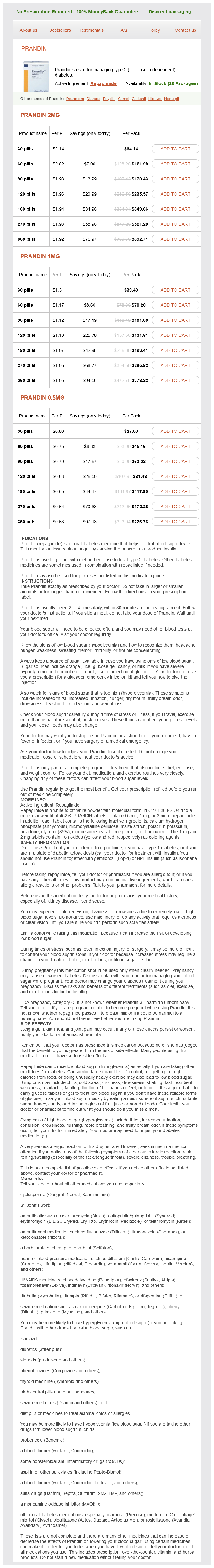
Prandin Dosage and Price
Prandin 2mg
- 30 pills - $64.14
- 60 pills - $121.28
- 90 pills - $178.43
- 120 pills - $235.57
- 180 pills - $349.86
- 270 pills - $521.28
- 360 pills - $692.71
Prandin 1mg
- 30 pills - $39.40
- 60 pills - $70.20
- 90 pills - $101.00
- 120 pills - $131.81
- 180 pills - $193.41
- 270 pills - $285.82
- 360 pills - $378.22
Prandin 0.5mg
- 30 pills - $27.00
- 60 pills - $45.16
- 90 pills - $63.32
- 120 pills - $81.48
- 180 pills - $117.80
- 270 pills - $172.28
- 360 pills - $226.76
Therefore diabetes y alcohol buy prandin 1 mg online, an advantage of polyurethane in certain designs is "higher stiffness" combined with "higher tear strength. In addition, some believe that polyurethane is inherently less thrombogenic than silicone rubber. Although silicone rubber has been available longer, polyurethane has been used in humans as lead insulation for >30 years. Polyurethane has been available in a softer, more flexible version known as "80A" and a harder, less flexible version known as "55D. Lead diameter In general, lead diameters have decreased over the years, and though this is of benefit for ease of vascular access, avoidance of tricuspid regurgitation, and subclavian vein stenosis, lead failure with very small leads is a potential disadvantage. It may be advantageous to use preferentially a smalldiameter lead in children and in patients with existing transvenous leads in whom the vessel lumen is compromised. More recently, a lumenless pacemaker lead (Select Secure lead model 3830, Medtronic, Inc. The lead is delivered using a current polyurethane leads utilize the 55D version insulation, which is much less prone to the degradation. Even though mineralization of encapsulating sheaths (extrinsic mineralization) is common, there is no evi dence that it causes lead insulation failure (although it greatly hinders removal of old leads). Mineralization of the silicone rubber per se (intrinsic mineralization) has also been rarely observed, but lead failures from this mechanism have been very rare. Polyurethane, being "relatively stiffer," is often used advantageously, especially in a portion or certain segment of a lead, and allows manufacturers to make smaller, tough leads that can have greater torquability, resulting in easier implantability. At one time, not being "repairable" was a disadvan tage for polyurethane insulated leads versus silicone leads. In the earlier years of cardiac pacing, experienced individuals would at times attempt outer insulation repair of silicone rubber insulated leads with medical adhesive and silicone film. Also, terminal pin replacement on unipolar leads could be performed, if necessary, with specific repair "kits" from the manufacturers. If a lead is malfunctioning or grossly damaged, it should be abandoned and capped, or extracted and replaced. Although no longer a significant issue, "sensitivity to manufacturing process" can also be a disadvantage for both materials. The potential for environmental surface cracking remains true for the 80A polyurethane, but it is not a significant mechanism of clinical failure in con temporary 55D polyurethane leads. The potential for metal ion oxidation remains an issue for contemporary 55D polyurethane insulated leads. Despite a blemish on polyurethane leads due to a high failure rate in the 1980s of the 80A version of polyurethane, the overall survival rate for other, newer polyurethane leads utilizing the 55D version has been excellent. Ideally, leads would never fail, but this level of reliability will never be reached. The acceptable failure rate for permanent pacing and defibrillation leads (and pulse generators for that matter) is a matter of continued debate. Instead, the lead is advanced through a deflectable sheath that is maneuvered to the specific site of interest. The lead is then passed through the sheath and the helix advanced into the myocardium at that site. Note that in the portion of the lead that will be positioned within the atrium, there are two ring electrodes (arrows). Sensing occurs via these "floating" atrial electrodes and allows Psynchronous pacing with a single lead. Choosing the Device Generator and Leads deflectable sheath (Medtronic C304) or fixedcurve sheath (Medtronic C315His sheath, specifically designed for parahisian pacing) without the use of a stylet. Implantation of this lead has been shown to be feasible, and more commonly used for physiological/His bundle pacing; longterm experience with the lead is still limited. Compatibility of lead and pulse generator the pacemaker lead or leads and pulse generator selected do not need to be from the same manufacturer. It is generally acceptable to "mix and match" leads from company X with a pulse generator from company Y, assuming similar functionality. What is mandatory is that the lead connector be compatible with the connec tor cavity of the pulse generator. Historically, unipolar leads were 5 or 6 mm in diam eter, and bipolar leads were of the bifurcated design with similar 5 or 6 mm sizes. In 1986, a voluntary standard for lead con nectors and connector cavities was established. This voluntary standard for leads and connectors incorpo rated sealing rings on a 3. Epicardial leads Epicardial pacing may still be necessary in patients with congenital cardiac anomalies that prevent the access needed for transvenous leads, with a prosthetic tricus pid valve, or with other tricuspid valve abnormalities that preclude lead placement across the valve. Epicardial leads have historically had higher pacing thresholds and less mechanical reli ability than transvenous leads. Platinized and steroid eluting epicardial leads are used in an attempt to keep epicardial pacing thresholds lower. As new leads become available, it is important to be aware of resources of lead survival and performance. Manufacturers are required by law to collect post market surveillance data on hardware performance. Various manufacturers use different approaches for col lection and analysis of this information, but perfor mance data should be available from any manufacturer on request. Followup data may be obtained from active registry information25,26 or from centers that publish survival and performance data on individual leads see Chapter 10 (Troubleshooting). A literature search is likely to yield implanters with information on many of the most widely used leads.
© 2025 Adrive Pharma, All Rights Reserved..