General Information about Oxcarbazepine
Trileptal works by blocking voltage-sensitive sodium channels in the mind, stopping abnormal electrical exercise from spreading and inflicting seizures. It is primarily used for treating partial seizures, which are seizures that originate from a selected area of the mind. Trileptal can also be prescribed for generalized seizures, which affect either side of the brain without delay.
Oxcarbazepine, also known as Trileptal, is a drugs used to treat seizures in patients with epilepsy. It belongs to a class of drugs often recognized as anticonvulsants and works by reducing irregular electrical activity in the mind. Trileptal has been a widely used and efficient therapy possibility for epilepsy since its approval by the Food and Drug Administration (FDA) in 2000.
In conclusion, Trileptal has been a priceless treatment possibility for patients with epilepsy for over two decades. Its comfort, low potential for drug interactions, and comparatively gentle side effects make it a popular alternative among healthcare providers and patients alike. Other seizure problems, corresponding to trigeminal neuralgia, have additionally shown positive responses to treatment with Trileptal. With proper monitoring and communication with a healthcare supplier, Trileptal can help individuals with epilepsy effectively manage their condition and enhance their high quality of life.
One of some nice advantages of Trileptal is its convenience. It is on the market in pill kind, making it easy to take and administer. Trileptal additionally has a protracted half-life, that means that it remains in the physique for an extended period, allowing for much less frequent dosing. This is beneficial for sufferers who could have hassle with strict medication schedules.
Before starting Trileptal, sufferers ought to inform their healthcare supplier of some other drugs they're taking, including over-the-counter medication and supplements. This will help to forestall any potential drug interactions. Trileptal is also not beneficial for sufferers with a historical past of bone marrow suppression or hypersensitivity to carbamazepine, as there could also be an elevated danger of serious side effects.
Epilepsy is a neurological dysfunction that is characterized by recurrent seizures. Seizures are attributable to sudden and irregular electrical activity in the brain, which can lead to symptoms corresponding to convulsions, loss of consciousness, and uncontrolled movements of the physique. Epilepsy affects roughly 3 million individuals within the United States alone and may have a major impression on a person's high quality of life.
Occasionally, Trileptal could trigger a critical side impact called Stevens-Johnson syndrome, a uncommon however potentially life-threatening pores and skin response. Symptoms embody a rash, blisters, and peeling skin. Patients experiencing these symptoms ought to search medical attention instantly.
Another advantage of Trileptal is its relatively low danger for drug interactions. Unlike some other anticonvulsants, it doesn't work together with oral contraceptives, making it a protected option for women of childbearing age. It also has a decrease incidence of side effects in comparison with other anticonvulsants, similar to dizziness, sedation, and memory impairment.
Trileptal is mostly well-tolerated by most patients, with the most typical unwanted facet effects being dizziness and drowsiness. These effects are sometimes mild and have a tendency to lower over time because the body adjusts to the medication. Less frequent unwanted aspect effects may embody imaginative and prescient adjustments, nausea, and double vision, which must be reported to a healthcare supplier.
Treatment with low-dose glucocorticoids relieves bone pain and may reverse the abnormal bone formation symptoms depression oxcarbazepine 150 mg purchase with visa. Intermittent bisphosphonate therapy has produced clinical improvement in a limited number of patients. Following transplantation, the marrow contains progenitor cells and normally functioning osteoclasts. Limited studies in small numbers of patients have suggested variable benefits following treatment with interferon -1, 1,25-dihydroxyvitamin D (which stimulates osteoclasts directly), methylprednisolone, and a low-calcium/highphosphate diet. Surgical intervention is indicated to decompress optic or auditory nerve compression. Orthopedic management is required for the surgical treatment of fractures and their complications including malunion and postfracture deformity. The major manifestations are due to narrowed cranial foramens with neural compressions that may result in optic atrophy, facial paralysis, and deafness. Endosteal hyperostosis with syndactyly, known as sclerosteosis, is a more severe form. The major manifestation is progressive linear hyperostosis in one or more bones of one limb, usually a lower extremity. The name comes 2964 from the radiographic appearance of the involved bone, which resem- bles melted wax that has dripped down a candle. Symptoms appear during childhood as pain or stiffness in the area of sclerotic bone. There may be associated ectopic soft tissue masses, composed of cartilage or osseous tissue, and skin changes overlying the involved bone, consisting of scleroderma-like areas and hypertrichosis. The literal translation of osteopoikilosis is "spotted bones"; it is a benign autosomal dominant condition in which numerous small, variably shaped (usually round or oval) foci of bony sclerosis are seen in the epiphyses and adjacent metaphyses. The main differentiating points are that bony lesions of osteopoikilosis are stable over time and do not accumulate radionucleotide on bone scanning. In some kindred, osteopoikilosis is associated with connective tissue nevi known as dermatofibrosis lenticularis disseminata, also known as Buschke-Ollendorff syndrome. Histologic inspection reveals thickened but otherwise normal trabeculae and islands of normal cortical bone. Bone biopsy and histomorphometry reveal increased rates of bone formation, decreased bone resorption with a marked decrease in osteoclasts, and dense lamellar bone. Empirical therapy includes pain control, and there may be beneficial response to bisphosphonate. Rickets causes delayed walking with waddling gait, short stature, and dolichocephalic skull with frontal bossing. Adult hypophosphatasia presents during middle age with painful, poorly healing metatarsal stress fractures or thigh pain due to femoral pseudofractures. It is important to recognize hypophosphatasia in adults because treatment with bisphosphonates can result in increased rather than decreased bone fragility. Serum parathyroid hormone, 25-hydroxyvitamin D, and 1,25-dihydroxyvitamin D levels are normal. In contrast to other forms of rickets and osteomalacia, calcium and vitamin D supplementation should be avoided because they may aggravate hypercalcemia and hypercalciuria. A low-calcium diet, glucocorticoids, and calcitonin have been used in a small number of patients with variable responses. Because fracture healing is poor, placement of intramedullary rods is best for acute fracture repair and for prophylactic prevention of fractures. The frequency of the severe neonatal and infantile forms is about 1 in 100,000 live births in Canada, where the disease is most common because of its high prevalence among Mennonites and Hutterites. The severity of the disease is remarkably variable, ranging from intrauterine death associated with profound skeletal hypomineralization at one extreme to premature tooth loss as the only manifestation in some adults. Severe cases are inherited in an autosomal recessive manner, but the genetic patterns are less clear for the milder forms. Perinatal hypophosphatasia becomes manifest during pregnancy and is often complicated by polyhydramnios and intrauterine death. The infantile form becomes clinically apparent before the age of 6 months with failure to thrive, rachitic deformities, functional craniosynostosis despite widely open fontanels (which are actually hypomineralized areas of the calvarium), raised intracranial pressure, and flail chest with predisposition to pneumonia. This is a rare disorder characterized by defective skeletal mineralization despite normal serum calcium and phosphate levels. Clinically, the disorder presents in middle-aged or elderly men with chronic axial skeletal discomfort. Radiographic findings are mainly osteosclerosis due to coarsened trabecular patterns typical of osteomalacia. It presents in both sexes; in middle age or later; and with progressive, intractable skeletal pain and fractures; worsening immobilization; and a debilitating course. Radiographic evaluation reveals generalized osteomalacia, osteopenia, and occasional pseudofractures. Histologic features include a tangled pattern of collagen fibrils with abundant osteoblasts and osteoclasts. As the postzygotic mutations occur at different stages of early development, the extent and type of tissue affected are variable and explain the mosaic pattern of skin and bone changes. Such mutations of the Gs proteincoupled receptor may cause autonomous function in bone (parathyroid hormone receptor); skin (melanocyte-stimulating hormone receptor); and various endocrine glands including ovary (follicle-stimulating hormone receptor), thyroid (thyroid-stimulating hormone receptor), adrenal (adrenocorticotropic hormone receptor), and pituitary (growth hormonereleasing hormone receptor). The skeletal lesions are composed largely of mesenchymal cells that do not differentiate into osteoblasts, resulting in the formation of imperfect bone. In some areas of bone, fibroblast-like cells develop features of osteoblasts in that they produce extracellular matrix that organizes into woven bone. In other areas, cells have features of chondrocytes and produce cartilage-like extracellular matrix.
The thyroid can be palpated with both hands from behind or while facing the patient medicine dictionary pill identification discount 600 mg oxcarbazepine free shipping, using the thumbs to palpate each lobe. It is best to use a combination of these methods, especially when nodules are small. After locating the cricoid cartilage, the isthmus, which is attached to the lower one-third of the thyroid lobes, can be identified and then followed laterally to locate either lobe (normally, the right lobe is slightly larger than the left). Features to be noted include thyroid size, consistency, nodularity, and any tenderness or fixation. An estimate of thyroid size (normally 1220 g) should be made, and a drawing is often the best way to record findings. Ultrasound imaging provides the most accurate measurement of thyroid volume and nodularity and is useful for assessment of goiter prevalence in iodine deficient regions. However, ultrasound is not indicated if the thyroid physical examination is normal. A bruit or thrill over the gland, located over the insertion of the superior and inferior thyroid arteries (supero- or inferolaterally), indicates increased vascularity, associated with turbulent rather than laminar blood flow, as occurs in hyperthyroidism. If the lower borders of the thyroid lobes are not clearly felt, a goiter may be retrosternal. With any central mass above the thyroid, the tongue should be extended, as thyroglossal cysts then move upward. The thyroid examination is not complete without assessment for lymphadenopathy in the supraclavicular and cervical regions of the neck. T4 and T3 are highly protein-bound, and numerous factors (illness, medications, genetic factors) can influence protein binding. It is useful, therefore, to measure the free, or unbound, hormone levels, which correspond to the biologically available hormone pool. Two direct methods are used to measure unbound thyroid hormones: (1) unbound thyroid hormone competition with radiolabeled T4 (or an analogue) for binding to a solid-phase antibody, and (2) physical separation of the unbound hormone fraction by ultracentrifugation or equilibrium dialysis. Although early unbound hormone immunoassays suffered from artifacts, newer assays correlate well with the results of the more technically demanding and expensive physical separation methods. The latter is derived from the T3-resin uptake test, which determines the distribution of radiolabeled T3 between an absorbent resin and the unoccupied thyroid hormone binding proteins in the sample. The binding of the labeled T3 to the resin is increased when there is reduced unoccupied protein binding sites. In effect, the index corrects for anomalous total hormone values caused by variations in hormone-protein binding. Because unbound thyroid hormone levels are normal and the patient is euthyroid in all of these circumstances, assays that measure unbound hormone are preferable to those for total thyroid hormones. For most purposes, the unbound T4 level is sufficient to confirm thyrotoxicosis, but 25% of patients have only an elevated T3 level (T3 toxicosis). Tests for the end-organ effects of thyroid hormone excess or depletion, such as estimation of basal metabolic rate, tendon reflex relaxation rates, or serum cholesterol, are relatively insensitive and are not useful as clinical determinants of thyroid function. About 515% of euthyroid women and up to 2% of euthyroid men have thyroid antibodies; such individuals are at increased risk of developing thyroid dysfunction. Serum Tg levels are increased in all types of thyrotoxicosis except thyrotoxicosis factitia caused by self-administration of thyroid hormone. Tg levels are particularly increased in thyroiditis, reflecting thyroid tissue destruction and release of Tg. The main role for Tg measurement, however, is in the follow-up of thyroid cancer patients. After total 2698 thyroidectomy and radioablation, Tg levels should be undetectable; in the absence of anti-Tg antibodies, measurable levels indicate incomplete ablation or recurrent cancer. Radioiodine Uptake and Thyroid Scanning the thyroid gland selectively transports radioisotopes of iodine (123I, 125I, 131I) and 99m Tc pertechnetate, allowing thyroid imaging and quantitation of radioactive tracer fractional uptake. Toxic adenomas appear as focal areas of increased uptake, with suppressed tracer uptake in the remainder of the gland. The vast majority of thyroid nodules do not produce thyroid hormone ("cold" nodules), and these are more likely to be malignant (~510%). Whole-body and thyroid scanning is also used in the treatment and surveillance of thyroid cancer. Thyroid Ultrasound Ultrasonography is valuable for the diagnosis and evaluation of patients with nodular thyroid disease (Chap. Evidence-based guidelines recommend thyroid ultrasonography for all patients suspected of having thyroid nodules by either physical examination or another imaging study. Sonographic patterns that combine suspicious sonographic features are highly suggestive of malignancy. In addition to evaluating thyroid nodules, ultrasound is useful for monitoring nodule size and for the aspiration of nodules or cystic lesions. Ultrasonography of the central and lateral cervical lymph node compartments is indispensable in the evaluation thyroid cancer patients, preoperatively and during follow-up. In addition, the American College of Radiology recommends a survey of the cervical lymph nodes as part of every diagnostic thyroid sonographic examination. Mutations that cause congenital hypothyroidism are being increasingly identified, but most remain idiopathic (Table 376-2). Transplacental passage of maternal thyroid hormone occurs before the fetal thyroid gland begins to function and provides partial hormone support to a fetus with congenital hypothyroidism. Although some patients may have minor symptoms, this state is called subclinical hypothyroidism. Prevalence the mean annual incidence rate of autoimmune hypo- thyroidism is up to 4 per 1000 women and 1 per 1000 men.
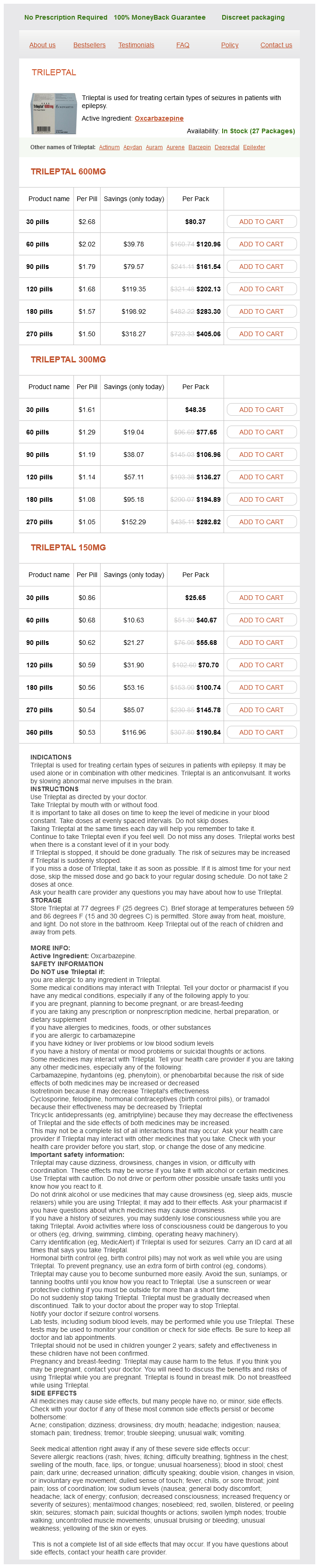
Oxcarbazepine Dosage and Price
Trileptal 600mg
- 30 pills - $80.37
- 60 pills - $120.96
- 90 pills - $161.54
- 120 pills - $202.13
- 180 pills - $283.30
- 270 pills - $405.06
Trileptal 300mg
- 30 pills - $48.35
- 60 pills - $77.65
- 90 pills - $106.96
- 120 pills - $136.27
- 180 pills - $194.89
- 270 pills - $282.82
Trileptal 150mg
- 30 pills - $25.65
- 60 pills - $40.67
- 90 pills - $55.68
- 120 pills - $70.70
- 180 pills - $100.74
- 270 pills - $145.78
- 360 pills - $190.84
Axial diffusion-weighted image (H) and apparent diffusion coefficient image (I) documents the presence of a right middle cerebral artery infarction treatment example oxcarbazepine 600 mg order line. Risk factors for contrast nephropathy include age (>80 years), preexisting renal disease (serum creatinine exceeding 2 mg/dL), solitary kidney, diabetes mellitus, dehydration, paraproteinemia, concurrent use of nephrotoxic medication or chemotherapeutic agents, and high contrast dose. Nonionic, low-osmolar media produce fewer abnormalities in renal blood flow and less endothelial cell damage but should still be used carefully in patients at risk for allergic reaction. In one study, 15% of outpatients with a normal serum creatinine had an estimated creatinine clearance of 50 mL/min/1. Use of other agents such as bicarbonate and acetylcysteine may reduce the incidence of contrast nephropathy. Subacute (>1 h after injection) reactions are frequent and probably related to T cellmediated immune reactions. Drug provocation and skin testing may be required to determine the culprit agent involved as well as determine a safe alternative. Extravasation of contrast media, although rare, can be painful and lead to compartment syndrome. Patients with significant cardiac disease may be at increased risk for contrast reactions, and in these patients, limits to the volume and osmolality of the contrast media should be considered. Patients who may undergo systemic radioactive iodine therapy for thyroid disease or cancer should not receive iodinated contrast media if possible, because this will decrease the uptake of the radioisotope into the tumor or thyroid (see the American College of Radiology Manual on Contrast Media, Version 10. Images are made by computerized processing of resonance information received from protons in the body. Spatial localization is achieved by magnetic gradients surrounding the main magnet, which impart slight changes in magnetic field throughout the imaging volume. Rf pulses transiently excite the energy state of the hydrogen protons in the body. The subsequent return to equilibrium energy state (relaxation) of the hydrogen protons results in a release of Rf energy (the echo), which is detected by the coils that delivered the Rf pulses. Allergy Immediate reactions following intravenous contrast media occur through several mechanisms. The most severe reactions are related to allergic hypersensitivity (anaphylaxis) and range from mild hives to bronchospasm and death. The pathogenesis of allergic hypersensitivity reactions is thought to include the release of mediators such as histamine, antibody-antigen reactions, and complement activation. Risk factors include a history of prior contrast reaction (fivefold increased likelihood), food and or drug allergies, and atopy (asthma and hay fever). The predictive value of specific allergies, such as those to shellfish, once thought important, actually is now recognized to be unreliable. If iodinated contrast is absolutely required, a nonionic agent should be used in conjunction with pretreatment with glucocorticoids and antihistamines (Table 416-2); however, pretreatment does not guarantee safety. It would be wise T1 and T2 Relaxation Times the rate of return to equilibrium of perturbed protons is called the relaxation rate. The relaxation rate of a hydrogen proton in a tissue is influenced by local interactions with surrounding molecules and atomic neighbors. Fat and subacute hemorrhage have relatively shorter T1 relaxation rates and thus higher signal intensity than brain on T1W images. T2W images are more sensitive than T1W images to edema, demyelination, infarction, and chronic hemorrhage, whereas T1W imaging is more sensitive to subacute hemorrhage and fat-containing structures. The selection of a proper protocol that will best answer a clinical question depends on an accurate clinical history and indication for the examination. This sequence interrogates the microscopic motion of water, which is restricted in areas of infarction, abscess, and some tumors. Each sequence, however, must be obtained separately and takes 110 min on average to complete. Coronal postcontrast T1-weighted image demonstrates a ring-enhancing mass in the left frontal lobe. Axial diffusion-weighted image demonstrates restricted diffusion (high signal intensity) within the lesion, which in this setting is highly suggestive of cerebral abscess. Gadolinium is a paramagnetic substance that reduces the T1 and T2 relaxation times of nearby water protons, resulting in a high signal on T1W images and a low signal on T2W images (the latter requires a sufficient local concentration, usually in the form of an intravenous bolus). These differ according to the attached chelated moiety, which also affects the strength of chelation of the otherwise toxic gadolinium element. The chelating carrier molecule for gadolinium can be classified by whether it is macrocyclic or has linear geometry and whether it is ionic or nonionic. The agents are generally well tolerated; overall adverse events after injection range from 0. Severe life-threatening reactions are exceedingly rare; in one report, only 55 reactions out of 20 million doses occurred. However, the adverse reaction rate in patients with a prior history of reaction to gadolinium is eight times higher than normal. There is no cross reactivity between different classes of contrast media; a prior reaction to gadolinium-based contrast does not predict a future reaction to iodinated contrast medium, or vice versa, more than any other unrelated allergy. Gadolinium contrast material can be administered safely to children as well as adults, although these agents are generally avoided in those aged <6 months. Coronal (A) and axial (B) T2-weighted fluidattenuated inversion recovery images demonstrate expansion and high signal intensity involving the right medial temporal lobe and insular cortex (arrows). Coronal diffusion-weighted image demonstrates high signal intensity indicating restricted diffusion involving the right medial temporal lobe and hippocampus (arrows) as well as subtle involvement of the left inferior temporal lobe (arrowhead). This is most consistent with neuronal death and can be seen in acute infarction as well as encephalitis and other inflammatory conditions. The suspected diagnosis of herpes simplex encephalitis was confirmed by cerebrospinal fluid polymerase chain reaction analysis. In addition to dermatologic symptoms, other manifestations include widespread fibrosis of the skeletal muscle, bone, lungs, pleura, pericardium, myocardium, kidney, muscle, bone, testes, and dura.
© 2025 Adrive Pharma, All Rights Reserved..