General Information about Levothroid
Levothroid, also called Levothyroxine, is a drugs used to deal with low ranges of thyroid hormones. The thyroid gland produces two primary hormones, thyroxine (T4) and triiodothyronine (T3), which play a vital role in regulating the physique's metabolism. When the thyroid gland just isn't functioning properly and is unable to supply sufficient hormones, it leads to a condition called hypothyroidism. Levothroid is an artificial form of T4 and is used to complement the physique's pure levels of thyroid hormones.
Hypothyroidism is a common condition that affects tens of millions of people worldwide. It is more frequent in women and people over the age of 60. The symptoms of hypothyroidism may be delicate and simply missed, making it difficult to diagnose. Some of the common indicators and signs embrace fatigue, weight gain, dry pores and skin, hair loss, muscle weak point, and sensitivity to chilly temperatures.
One of the advantages of Levothroid is its minimal unwanted effects. As it is a synthetic type of the natural thyroid hormone, it's well-tolerated by most individuals. However, like several treatment, some folks might experience unwanted side effects, which may embrace headache, insomnia, and modifications in appetite. In some rare cases, sufferers may expertise an allergic reaction to Levothroid, which may manifest as hives, problem breathing, and swelling of the face and tongue. In such instances, instant medical consideration should be sought.
Levothroid works by replacing the poor thyroid hormones within the physique. It is out there in tablet type and may be taken orally, normally once a day. The dosage could differ relying on the individual's age, weight, and severity of hypothyroidism. It is important to take Levothroid at the identical time every day to maintain a consistent stage of thyroid hormones in the body.
In conclusion, Levothroid is a life-saving medication for people with hypothyroidism. It is a safe and efficient method to complement the body's natural levels of thyroid hormones. However, it's crucial to take the medication as prescribed and inform your doctor about some other drugs or supplements you are taking. With correct therapy, people with hypothyroidism can lead a wholesome and fulfilling life.
Levothroid can additionally be protected to make use of during being pregnant. In reality, untreated hypothyroidism throughout being pregnant can result in critical complications, similar to low birth weight, preterm birth, and neurodevelopmental points in the child. Therefore, it is essential for pregnant women with hypothyroidism to take Levothroid as prescribed by their physician to make sure a healthy being pregnant.
It is crucial to notice that Levothroid is not a cure for hypothyroidism. It is a lifelong treatment that helps manage the condition and keep the physique's metabolism. Therefore, it is essential for patients to proceed taking Levothroid even when their signs enhance. Suddenly stopping the medication or altering the dosage without consulting a doctor can lead to a relapse or different complications.
Levothroid might work together with different drugs, dietary supplements, or foods, which may affect the absorption and effectiveness of the medication. Therefore, it is important to inform your doctor about some other medications or dietary supplements you are taking before beginning Levothroid. It can be really helpful to take Levothroid on an empty stomach, a minimum of half-hour earlier than breakfast or any other drugs.
Antimicrobial proteins and peptides are present in neutrophil cytoplasmic granules thyroid symptoms worse at night order 100 mcg levothroid amex. Other granule components, such as Chapter 7: Hematology of the Fetus and Newborn 107 myeloperoxidase (bacterial killing) and defensins (antimicrobial proteins), are not diminished. Cytokine Effects on Neonatal Phagocytic Function There is a complex interaction between cytokines produced by lymphocytes and macrophages, and the activation status of neutrophils during infection. Low levels of this receptor also impair complement-mediated phagocytosis and oxidative metabolism. The bleeding time reflects platelet function and capillary integrity, as well as the platelet count, and traditionally has been used to assess these parameters. However, there are technical difficulties in applying a technique for measuring bleeding time to neonates or preterm infants because of the need for venous occlusion of the forearm, where the test Platelet Functions normally is performed, and for a minimal incision to avoid scarring of the skin. Bleeding times were measured using an automatic device to minimize trauma in normal neonates, with venous occlusion of 20 torr for infants who weigh less than 1000 g, 25 torr for those who weigh 1000 to 2000 g, and 30 torr for those who weigh more than 2000 g. The result of these differences is shortened bleeding and closure times in normal neonates (see below). The use of indomethacin for treatment of patent ductus arteriosus in preterm infants has been questioned because this agent interferes with prostaglandin metabolism and the production of thromboxane A2, an important initiator of platelet aggregation. The closure time to assess platelet function may replace the bleeding time, particularly for neonates and young children in whom bleeding times are difficult to perform and interpret. Newborn infants have closure times that are shorter than those of adults, likely related to their higher hematocrits, increased von Willebrand multimers and hence ristocetin cofactor, and higher leukocyte counts. Platelet Aggregation and Metabolism A variety of differences have been described in the platelet function of neonates. The in vitro findings do not appear related to a significant defect in prostaglandin synthesis or to storage pool deficiency of adenine nucleotides. These in vitro abnormalities may aggravate the impairment in platelet function and the predisposition to bleeding that result from neonatal diseases, particularly respiratory distress syndrome and sepsis. Maternal aspirin ingestion also results in abnormalities in platelet aggregation in the newborn in response to collagen. They are presumably caused by trauma associated with passage through the birth canal and disappear within a few days. Approximately 2 percent of the population in the United States of European descent is homozygous for PlA2 and thus are PlA1-negative. The complete expression of the PlA1 antigen during early gestation likely permits sensitization in women who are PlA1-negative even during their first pregnancy. Blood Lymphocyte Subsets: Infants Age 1 to 3 Days Median (10th90th percentile range) Lymphocyte Subsets Lymphocytes × 10 /L 9 Infants (13 Days) 3. Humoral (B-cell) immunity also develops early in gestation,236 but it is not fully active until after birth. In the newborn, approximately 15 percent of lymphocytes have immunoglobulin on their surface, with all immunoglobulin isotypes represented. The percentages of B cells expressing specific immunoglobulin isotypes are not related to the plasma levels of those isotypes. Variation in antibody response to specific antigens relates to the interaction of macrophages, T cells, and B cells. B lymphocytes are well represented in newborns, but T-lymphocyte independent B-lymphocyte responses are limited during the first year. Animals kept germ-free after birth have few plasma cells and markedly decreased production of immunoglobulins. Breastfeeding provides some transfer of antibodies, particularly secretory IgA, lysozyme, and lactoferrin. Large numbers of lymphocytes and monocytes (106 cells/mL) are found in colostrum and milk during the first 2 months postpartum. Although the newborn infant can produce specific IgG antibody,263 only small amounts of IgG are usually produced by the fetus. IgG levels in premature infants are reduced in relation to gestational age because of the low placental transport early in pregnancy. These differences from the adult may relate to functional immaturity of B and T lymphocytes,269271 to increased activity of suppressor T cells,258,269 and perhaps to altered macrophage function. These "pocks" represent residual intraerythrocyte inclusions, which remain because of monocyte and macrophage hypofunction. In spite of the lower levels of factors, the functional tests (prothrombin and partial thromboplastin times) are only slightly prolonged compared to adult normal values Table 76). Although different coagulation factors show different postnatal patterns of maturation, near-adult values are achieved for most components by 6 months of age. This fall may be lessened by administration of vitamin K,286 effectively preventing classic, early occurring (first few days after birth) hemorrhagic disease of the newborn. Inactive prothrombin molecules have been found in the plasma of some newborns, but they disappear after administration of vitamin K. A hemorrhagic diathesis also may occur later, 2 to 12 weeks after birth, as a result of lack of vitamin K, and is called late hemorrhagic disease of the newborn or acquired prothrombin complex deficiency. This problem can be prevented by parenteral or oral vitamin K, but the preferred route of administration remains controversial. The range of values encompassing 95 percent of the population is shown in parentheses. Data from Andrew M, Paes B, Milner B, et al: Development of the human coagulation system in the full-term infant. Chapter 7: Hematology of the Fetus and Newborn 111 parenteral route may result rarely in neuromuscular complications,293 and an association of intramuscular vitamin K prophylaxis and cancer in infancy was suggested but not substantiated. Oral administration, however, appears less reliable and may require repeated doses.
From the hematologic standpoint thyroid gland structure 100 mcg levothroid order amex, it is convenient to divide the causes of B12 deficiency into those that frequently lead to megaloblastic anemia and those that usually do not. Many other causes of defective cobalamin absorption involve mainly the stomach, or small intestine and to lesser extent, the pancreas. The term pernicious anemia sometimes is used as a synonym for cobalamin deficiency, but it should be reserved for the condition resulting from defective secretion of intrinsic factor by an atrophic gastric mucosa caused by an autoimmune process primarily directed against the parietal cells and their products. The disease is associated with human leukocyte antigen types A2, A3, B7, and B12,275 and with blood group A. In Americans of African descent, the disease tends to begin early, occurs with high frequency in women, and often is severe. There is an approximately twofold increase in the incidence of gastric cancer, similar increases in the incidence of certain hematologic malignancies, and an increase in the incidence of gastric carcinoid. Cells recovered by lavage are large168 and show atypical nuclei resembling early malignant change. The disease is easily missed because of its (1) insidious onset, (2) tendency to be masked by the use of multivitamin preparations containing folic acid,292 and (3) many atypical presentations,293 including its presentation as a neurologic disease without hematologic findings,77,294 and its tendency to be overlooked in patients with another autoimmune disease. Antiparietal cell and antiintrinsic factor antibodies are rarely measured, even though antiintrinsic factor antibodies in particular could be of considerable diagnostic value. The thick mucosa is packed with gastric glands composed mostly of chief cells and parietal cells. Gastric glands in the atrophic mucosa are sparse and consist mainly of mucus-secreting cells. Iron-deficiency anemia is most common, but cobalamin deficiency with megaloblastic anemia can occur. After total gastrectomy, cobalamin deficiency develops within 5 or 6 years because the operation removes the source of intrinsic factor. This may occur more rapidly because of abrogation of the enterohepatic reabsorption of biliary cobalamin. After partial gastrectomy, few patients show frank cobalamin deficiency, but approximately 5 percent have intermediate megaloblastosis, approximately 25 to 50 percent have low serum cobalamin levels, and many have varying degrees of decreased cobalamin absorption. Postgastrectomy patients with low serum cobalamin levels usually have low serum iron levels,297 in contrast to the high iron levels otherwise typical of cobalamin deficiency. A surgical procedure that has gained popularity for the treatment of morbid obesity is gastric reduction surgery. This procedure results in multiple deficiencies of micronutrients including cobalamin. Competing Intestinal Flora and Fauna: "Blind Loop Syndrome" the blind loop syndrome is a state of cobalamin malabsorption with megaloblastic anemia caused by intestinal stasis from anatomic lesions (strictures, diverticula, anastomoses, surgical blind loops) or impaired motility (scleroderma, amyloid). Cobalamin malabsorption is not corrected by exogenous intrinsic factor but may be corrected by antibiotic treatment. The defect in cobalamin absorption is caused by colonization of the diseased small intestine by bacteria that take up ingested cobalamin before it can be absorbed from the intestine. Another cause of cobalamin deficiency is infestation with the fish tapeworm Diphyllobothrium latum. Prevalence is highest near the Baltic Sea, Canada, and Alaska, where raw or undercooked fish is consumed. Cobalamin deficiency results from competition between the worm and the host for ingested cobalamin. The onset of cobalamin deficiency in vegans is slower than in conditions associated with cobalamin malabsorption. Thus it may take 10 to 20 years for an individual consuming a vegan diet to manifest features of cobalamin deficiency. In addition to vegans, however, there is mounting evidence of cobalamin inadequacy in children and young adults in developing countries that cannot be explained on the basis of cobalamin malabsorption, and has therefore been attributed to inadequate dietary intake. A megaloblastic anemia not related to cobalamin deficiency may accompany kwashiorkor or marasmus. These include a possible increase in breast cancer risk in premenopausal women330 and in osteoporosis. Cobalamin deficiency causes a neurologic syndrome that is particularly dangerous because the syndrome can develop in isolation,333 with no megaloblastic anemia to suggest a lack of cobalamin,294,334 and because the syndrome cannot be reversed by treatment when it is sufficiently far advanced. The syndrome usually begins with paresthesias in feet and fingers as a result of early peripheral neuropathy and disturbances of vibratory sense and proprioception. The earliest signs, which precede other neurologic findings by months, are loss of position sense in the second toe and loss of vibration sense for a 256-Hz but not a 128-Hz tuning fork. Somnolence and perversion of taste, smell, and vision with occasional optic atrophy are accompanied by slow waves on the electroencephalogram. Neurologic disorders closely resembling combined system disease develop in cobalamin-deficient fruit bats,324 pigs, and monkeys. Serum cobalamin may be normal, borderline, or low, but tissue cobalamin deficiency is suggested by consistently high levels of serum methylmalonic acid and/or homocysteine. Most of the neuropsychiatric abnormalities appear to respond to cobalamin therapy. Patients deficient in both cobalamin and folate may show normal serum folate levels. Plasma or Serum Cobalamin Levels intestinal bacteria synthesize propionate, a precursor of methylmalonate, and in conditions of bacterial overgrowth, microbially derived methylmalonic acid may contribute to elevations in plasma methylmalonic acid.
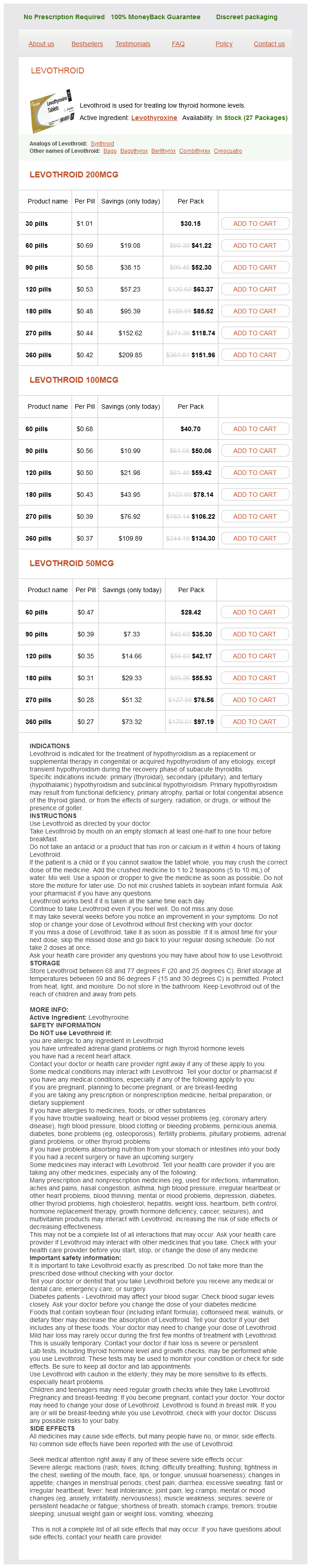
Levothroid Dosage and Price
Levothroid 200mcg
- 30 pills - $30.15
- 60 pills - $41.22
- 90 pills - $52.30
- 120 pills - $63.37
- 180 pills - $85.52
- 270 pills - $118.74
- 360 pills - $151.96
Levothroid 100mcg
- 60 pills - $40.70
- 90 pills - $50.06
- 120 pills - $59.42
- 180 pills - $78.14
- 270 pills - $106.22
- 360 pills - $134.30
Levothroid 50mcg
- 60 pills - $28.42
- 90 pills - $35.30
- 120 pills - $42.17
- 180 pills - $55.93
- 270 pills - $76.56
- 360 pills - $97.19
Ogata K thyroid cancer rates by state generic levothroid 200 mcg free shipping, Mendell-Harary J, Tachibana M, et al: Clinical safety, tolerability, pharmacokinetics, and pharmacodynamics of the novel factor Xa inhibitor edoxaban in healthy volunteers. Effectiveness of intravenous thrombolytic treatment in acute myocardial infarction. Jessup M, Antman E: Reducing the risk of heart attack and stroke: the American Heart Association/American College of Cardiology prevention guidelines. Huang Y, Cheng Y, Wu J, et al: Cilostazol as an alternative to aspirin after ischaemic stroke: A randomised, double-blind, pilot study. Jull A, Arroll B, Parag V, Waters J: Pentoxifylline for treating venous leg ulcers. Cosmi B, Rubboli A, Castelvetri C, Milandri M: Ticlopidine versus oral anticoagulation for coronary stenting. Valgimigli M, Biondi-Zoccai G, Tebaldi M, et al: Tirofiban as adjunctive therapy for acute coronary syndromes and percutaneous coronary intervention: A meta-analysis of randomized trials. Tricoci P, Huang Z, Held C, et al: Thrombin-receptor antagonist vorapaxar in acute coronary syndromes. Adoptive T-cell therapy, in which T cells are isolated or engineered to be specific for molecules expressed on diseased cells and administered to patients, has shown efficacy in infections and malignancy. Clinical applications of T-cell therapy have been facilitated by identification of target antigens expressed by viruses and tumors, improvement in strategies for the isolation and genetic engineering of antigen-specific T cells with intrinsic qualities that enable their persistence in vivo, and recognition that transferring T cells into a lymphopenic environment improves the efficiency of cell transfer and treatment efficacy. Insights into the obstacles to routinely achieving an effective antitumor response either by T-cell therapy or vaccination have been derived from careful analysis of clinical trials, and further development of immune cell therapy combined with interventions that target specific regulatory or inhibitory pathways that are present in tumor microenvironments and impede effective immunity represent promising areas for future applications. However, the isolation and propagation of antigen-specific T-cell clones requires specific expertise and is time-consuming. Antigen-specific T cells can be isolated by in vitro culture, expanded in long- or short-term culture, and then transferred in large numbers to patients. Alternatively, antigen-specific T cells can be isolated using direct methods and transferred immediately to the patient at low numbers. Patients are monitored after each cell infusion for toxicity, T-cell persistence, and efficacy. Chapter 26: Immune Cell Therapy 411 Transferred T cells must persist as functional memory T cells and migrate to sites of virus replication to be effective. Therefore, it was essential to verify the presence of the transferred T cells in the blood based on functional or structural properties. These techniques are being employed to enumerate and analyze the function of cells on a single-cell level. The phenotype of naïve, memory, and effector subsets is shown and the linear pathway of differentiation from a naïve T cell is based on recent data from fate mapping studies in murine models. Classes of tumor cell surface molecules for chimeric antigen receptor-modified T cells · B-cell differentiation molecules. Antiviral drugs may benefit a subset of patients, but the effects are often limited and accompanied by toxicities. Adoptive transfer of these T cells to 11 allogeneic transplant recipients produced a greater than 90 percent sustained virologic and clinical response. However, the clinical translation of adoptive T-cell therapy and other immunotherapeutic modalities for human cancers has proven to be more challenging than for opportunistic viral infections. Melanoma has served as a model for the discovery of human tumor antigens because T cells specific for melanoma cells can often be detected in the blood or the tumor microenvironment. Mutated proteins that arise as a consequence of the genetic instability of tumors are being identified as critical targets of immune recognition, and these offer the greatest promise for selectively targeting tumor cells without recognition of normal cells. Studies of the mechanisms of tumor eradication in melanoma are providing insights for treatment of other cancers with T-cell therapy. Treatment with a greater than 95 percent pure population of mutation-reactive T cells resulted in dramatic and durable tumor regression in this patient. Advances in our understanding of the role of individual cytokines in T-cell survival in vitro and in vivo, or the regulation of T-cell activation and homeostasis will provide new opportunities for improving the persistence of in vitro expanded T cells after transfer, perhaps obviating use of toxic chemoradiotherapy to deplete lymphocytes before T-cell infusions. Two approaches that have already been translated to the clinic are discussed below. After isolation of T cells from the desired T-cell subset, the tumor targeting receptor is introduced in the T cells by gene transfer, and the engineered redirected T cells are expanded for reinfusion to the patient. Five patients experienced cancer regression, but three patients experienced serious and/or fatal neurologic toxicity. Suitable animal models for preclinical toxicity studies are urgently needed and are being developed. Advances in the understanding of cell intrinsic properties of T-cell subsets, discovery of target antigens that distinguish tumor cells from normal cells, and improvements in the methodology for introducing genes into T cells have combined to make it feasible to treat patients with certain malignancies using highly effective T-cell products. For most common human tumors, target antigens have not yet been defined, and tumor heterogeneity and other mechanisms that tumors use to evade T-cell recognition represent barriers to effective therapy. Combination therapies with T cells and checkpoint inhibitors are promising for overcoming local and systemic evasion mechanisms that limit antitumor immunity. Finally, it would be ideal if expression of the tumor targeting receptors or the survival of transferred T cells were under regulatory control by small molecules that could be administered to the patient to reduce toxicity. Klenerman P, Hill A: T cells and viral persistence: Lessons from diverse infections. Tomblyn M, Chiller T, Einsele H, et al: Guidelines for preventing infectious complications among hematopoietic cell transplantation recipients: A global perspective. Pollack M, Heugel J, Xie H, et al: An international comparison of current strategies to prevent herpesvirus and fungal infections in hematopoietic cell transplant recipients. Reusser P, Einsele H, Lee J, et al: Randomized multicenter trial of foscarnet versus ganciclovir for preemptive therapy of cytomegalovirus infection after allogeneic stem cell transplantation. The results of clinical trials of T-cell therapy have revealed toxicity to normal tissues in some patients. The introduction of a suicide gene into the T cells that could be activated if toxicity occurred has long been the subject of research.
© 2025 Adrive Pharma, All Rights Reserved..