General Information about Levonorgestrel Bp
Levonorgestrel BP, also called Alesse, is a extremely effective contraception technique that is taken orally to stop ovulation and pregnancy. It is an artificial form of the female hormone progesterone, and its major operate is to inhibit the release of an egg from the ovaries. Alesse is a popular contraceptive amongst women due to its ease of use, effectiveness, and minimal unwanted facet effects.
In conclusion, Levonorgestrel BP (Alesse) is a highly efficient and handy birth control method that's widely utilized by girls to prevent pregnancy. Its excessive effectiveness, ease of use, and minimal unwanted effects have made it a well-liked selection amongst ladies worldwide. However, like any medicine, it is essential to make use of Alesse as directed and consult with a healthcare professional to determine if it's the proper possibility for particular person wants. With the use of Alesse, ladies can have more management over their reproductive well being and make knowledgeable choices about their future.
It is essential to notice that Alesse may not be appropriate for everybody. Women who've a history of blood clots, heart illness, liver disease, or breast most cancers ought to consult their physician earlier than beginning Alesse. Additionally, the capsule will not be as effective for girls who're overweight or on certain drugs which will have an result on its absorption. It is all the time really helpful to seek the guidance of with a healthcare skilled to determine if Alesse is the right birth control technique for one's particular person wants.
Alesse has been approved by the FDA and is extensively available in many countries around the globe. It is considered a mix contraception capsule, because it incorporates both progestin (levonorgestrel) and estrogen (ethinyl estradiol). The hormones in Alesse work together to thicken the cervical mucus, making it difficult for sperm to achieve an egg, and likewise thinning the liner of the uterus to forestall implantation.
Alesse can additionally be known for its ease of use. Unlike different birth control methods such as condoms or diaphragms, which require cautious placement and possible interruption throughout sexual activity, Alesse only requires one capsule to be taken at the similar time every single day. This makes it a convenient and discreet choice for ladies who may not wish to interrupt intimacy for contraceptive functions.
Additionally, Alesse is understood to have minimal side effects in comparison with other contraception methods. Some girls might experience gentle side effects such as nausea, headache, or breast tenderness, but these usually subside within the first few months of taking the capsule. Alesse additionally has reported positive effects on menstrual intervals, corresponding to decreasing cramps and regulating the cycle.
One of the principle advantages of using Alesse is its excessive level of effectiveness. When taken correctly, it is estimated to have a 99% success price in preventing being pregnant. This means that out of every one hundred ladies utilizing Alesse, just one might become pregnant. However, it may be very important notice that Alesse does not defend towards sexually transmitted infections (STIs), and in some uncommon circumstances, when it is not taken accurately, it may not prevent pregnancy.
Hemorrhagic Fever Diseases A number of agents that infect the blood and lymphatics cause extreme fevers birth control pills necon 135 buy levonorgestrel canada, some of which are accompanied by internal hemorrhaging. Most of these viruses are zoonotic and their geographic pattern of distribution is determined by the presence of their natural hosts. Bunyaviridae is a family with members that cause hemorrhagic fevers, such as Rift Valley fever, which is endemic to Africa. Although we do not discuss examples of such diseases here, it is important to note that the prevalence of many of these diseases fluctuates today due to global warming patterns. Ebola and Marburg the Ebola and Marburg viruses are filoviruses (family Filoviridae). The two are related and cause similar symptoms, although Ebola has received the greatest share of media attention. Its gruesome symptoms are extreme manifestations of the same kind of hemorrhagic events described for dengue fever. The virus in the bloodstream leads to extensive capillary fragility and disruption of clotting. Patients bleed from their orifices, even from their mucous membranes, and experience massive internal and external hemorrhage. The mortality rate for Marburg infection is 25%, while it is a staggering 70% in cases of Ebola infection. Outbreaks with Marburg virus are rare, but individuals have been infected sporadically since it was first recognized in 1967. Dengue fever is also called "breakbone fever" because of the severe pain it can induce in muscles and joints (it does not actually cause fractures). The illness is endemic to Southeast Asia and India, and several epidemics have occurred in South America and Central America, the Caribbean, and Mexico. This may sound surprising, since health care workers can become infected even after wearing personal protective equipment. The people at most risk are those who are caring for patients in the late stages of disease, where there might be copious amounts of body fluids (blood, vomit, diarrhea). The virus In 2014, an Ebola epidemic began in West Africa, and it was destined to be the largest outbreak in history. Prior to that time, sporadic outbreaks had occurred in isolated regions, but in 2014 the conditions were just right. And as of April 2016, 28,646 suspected, probable, or confirmed cases had occurred, and 11,323 people had died. Guinea Sierra Leone Liberia In the table are the important features of the virus in more detail than in the text. Epidemiologists classify infectious agents according to two important factors: their likelihood of infecting contacts (infectivity or infectiousness) and their virulence (deadliness). In a firstworld country like the United States, health care workers are at highest risk. Among the first transmissions in the United States, an infected man traveling from Liberia was hospitalized in Dallas, and at least one of his nurses became infected, but she survived. Avoiding contact with patients and their fluids; two vaccines are in human trials Only supportive A fever of > 101. In Africa, a 55% fatality rate Occurred mainly in three African countries (Sierra Leone, Liberia, and Guinea) and was harder to control because it hit urban areas of dense population Possible transmission through semen Treatment Distinctive Features Epidemiology High fatality rate: 30%90% Occurs in sporadic outbreaks in Africa, often in rural areas where outbreaks can be contained due to limited population 20. Several related arenaviruses cause the diseases Argentine hemorrhagic fever, Bolivian hemorrhagic fever, and lymphocytic choriomeningitis (an infection of the brain and meninges). Lassa fever virus is found primarily in West Africa, but imported cases of disease have been identified in the United Kingdom. This means that although they became ill while in the United Kingdom, the patients acquired their actual infection while in Africa. In most cases, infection with this virus is asymptomatic, but in 20% of the cases a severe hemorrhagic syndrome develops. The syndrome includes chest pain, hemorrhaging, sore throat, back pain, vomiting, diarrhea, and sometimes encephalitis. The reservoir of the virus is a rodent found in sub-Saharan Africa called the multimammate rat. The virus is spread to humans through aerosolization of rat droppings, urine, hair, and so forth. Vertical transmission also occurs, and the disease leads to spontaneous abortions in 95% of infected pregnant women. This hemorrhagic fever has been shown to respond to the antiviral agent ribavirin, especially if administered in the early stages of infection. Because it is able to avoid destruction in the phagocytes, the bacterium is transported easily through the bloodstream and to various organs, such as the liver, kidney, breast tissue, or joints. Scientists suspect that the up-anddown nature of the fever is related to unusual properties of the bacterial lipopolysaccharide. Transmission and Epidemiology and Symptoms the Brucella species responsible for this disease live in phagocytic cells. These cells carry the bacteria into the bloodstream, creating focal lesions in the liver, spleen, bone marrow, and kidney. The cardinal manifestation of human brucellosis is a fluctuating pattern of fever, which is the origin of the common name undulant fever (figure 20. It is also accompanied by chills, profuse sweating, headache, muscle pain and weakness, and weight loss. Fatalities are not common, although the syndrome can last for a few weeks to a year, even with treatment. Causative Agent the bacterial genus Brucella contains tiny, aerobic gram-negative coccobacilli. Even though a principal manifestation of the disease in animals is an infection of the placenta and fetus, human placentas do not become infected.
Visceral movement associated with digestion occurs freely birth control pills you can take while breastfeeding discount levonorgestrel generic, and the doublelayered reflections of peritoneum passing between the walls and the viscera provide passage for the blood vessels, lymphatics, and nerves. Variable amounts of fat may also occur between the walls and viscera and the peritoneum lining them. The plane of the pelvic inlet (superior pelvic aperture) arbitrarily, but not physically, separates the abdominal and the pelvic cavities. Consequently, the more superiorly placed abdominal organs (spleen, liver, part of the kidneys, and stomach) are protected by the thoracic cage. The greater pelvis (expanded part of the pelvis superior to the pelvic inlet) supports and partly protects the lower abdominal viscera (part of the ileum, cecum, appendix, and sigmoid colon). The body has been sectioned in the median plane to show the abdominal and pelvic cavities as subdivisions of the continuous abdominopelvic cavity. Nine regions of the abdominal cavity are used to describe the location of abdominal organs, pains, or pathologies (Table 5. The regions are delineated by four planes: two sagittal (vertical) and two transverse (horizontal) planes. Some clinicians use the transpyloric and interspinous planes to establish the nine regions. Because the viscera sag with the pull of gravity, the pylorus usually lies at a lower level when the individual is standing erect. It is important to know what organs are located in each abdominal region or quadrant so that one knows where to auscultate, percuss, and palpate them (Table 5. The abdominal wall is musculo-aponeurotic, except for the posterior wall, which includes the lumbar region of the vertebral column. The boundary between the anterior and lateral walls is indefinite; therefore, the term anterolateral abdominal wall is often used. Some structures, such as muscles 976 and cutaneous nerves, are in both the anterior and lateral walls. A transverse section of the abdomen demonstrates various aspects of the wall and its components. The anterior abdominal wall and soft tissues of the anterior thoracic wall have been removed. Most of the intestine is covered by the apron-like greater omentum, a peritoneal fold hanging from the stomach. Layers of the anterolateral abdominal wall, including the trilaminar flat muscles, are shown. The skin attaches loosely to the subcutaneous tissue, except at the umbilicus, where it adheres firmly. Most of the anterolateral wall includes three musculotendinous layers; the fiber bundles of each layer run in different directions. This three-ply structure is similar to that of the intercostal spaces in the thorax. Fascia of Anterolateral Abdominal Wall the subcutaneous tissue over most of the wall includes a variable amount of fat. Males are especially susceptible to subcutaneous accumulation of fat in the lower anterior abdominal wall. In morbid obesity, the fat is many inches thick, often forming one or more sagging folds (L. Superior to the umbilicus, the subcutaneous tissue is consistent with that found in most regions. Inferior to the umbilicus, the deepest part of the subcutaneous tissue is reinforced by many elastic and collagen fibers, so it has two layers: the superficial fatty layer (Camper fascia) and the deep membranous layer (Scarpa fascia) of subcutaneous tissue. Superficial, intermediate, and deep layers of investing fascia cover the external aspects of the three muscle layers of the anterolateral abdominal wall and their aponeuroses (flat expanded tendons) and cannot be easily separated from them. The investing fascias here are extremely thin, being represented mostly by the epimysium (outer fibrous connective tissue layer surrounding all 979 muscles-see Chapter 1, Overview and Basic Concepts) superficial to or between muscles. The internal aspect of the abdominal wall is lined with membranous and areolar sheets of varying thickness constituting endoabdominal fascia. Although continuous, different parts of this fascia are named according to the muscle or aponeurosis it is lining. The portion lining the deep surface of the transversus abdominis muscle and its aponeurosis is the transversalis fascia. The glistening lining of the abdominal cavity, the parietal peritoneum, is formed by a single layer of epithelial cells and supporting connective tissue. The parietal peritoneum is internal to the transversalis fascia and is separated from it by a variable amount of extraperitoneal fat. The three flat muscles are the external oblique, internal oblique, and transversus abdominis. The muscle fibers of these three concentric muscle layers have varying orientations, with the fibers of the outer two layers running diagonally and perpendicular to each other for the main part, and the fibers of the deep layer running transversely. The aponeuroses then interweave with their fellows of the opposite side, forming a midline raphe (G. The decussation and interweaving of the aponeurotic fibers here is not only between right and left sides but also between superficial and intermediate and intermediate and deep layers. Intramuscular and intermuscular fiber exchanges within the bilaminar aponeuroses of the external and internal oblique muscles are shown.
Levonorgestrel Bp Dosage and Price
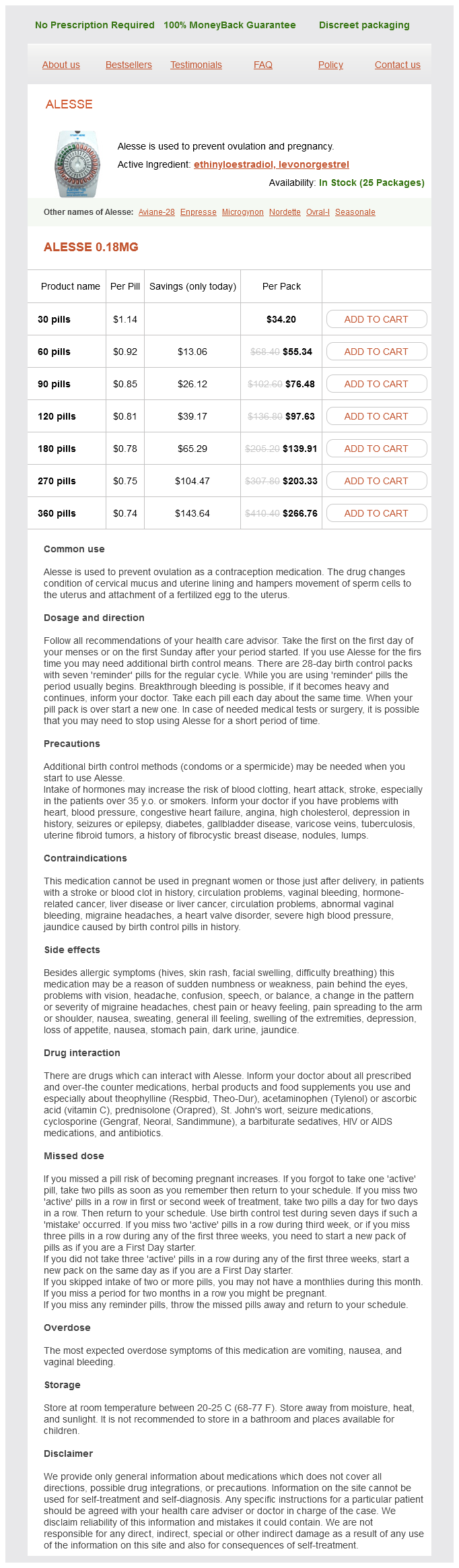
Alesse 0.18mg
- 30 pills - $34.20
- 60 pills - $55.34
- 90 pills - $76.48
- 120 pills - $97.63
- 180 pills - $139.91
- 270 pills - $203.33
- 360 pills - $266.76
The muscular system (myology) consists of skeletal muscles that act (contract) to move or position parts of the body birth control pills 84 days purchase 0.18mg levonorgestrel amex. The nervous system (neurology) consists of the central nervous system (brain and spinal cord) and the peripheral nervous system (nerves and ganglia, together with their motor and sensory endings). The sense organs, including the olfactory organ (sense of smell), eye or visual system (ophthalmology), ear (sense of hearing and balance-otology), and gustatory organ (sense of taste), are often considered with the nervous system in systemic anatomy. The cardiovascular system (cardiology) consists of the heart and blood vessels that propel and conduct blood through the body, delivering oxygen, nutrients, and hormones to cells and removing their waste products. The alimentary or digestive system (gastroenterology) consists of the digestive tract from the mouth to the anus, with all its associated organs and glands that function in ingestion, mastication (chewing), deglutition (swallowing), digestion, and absorption of food and the elimination of the solid waste (feces) remaining after the nutrients have been absorbed. The respiratory system (pulmonology) consists of the air passages and lungs that supply oxygen to the blood for cellular respiration and eliminate carbon 88 dioxide from it. The diaphragm and larynx control the flow of air through the system, which may also produce tone in the larynx that is further modified by the tongue, teeth, and lips into speech. The urinary system (urology) consists of the kidneys, ureters, urinary bladder, and urethra, which filter blood and subsequently produce, transport, store, and intermittently excrete urine (liquid waste). The genital (reproductive) system (gynecology for females; andrology for males) consists of the gonads (ovaries and testes) that produce oocytes (eggs) and sperms, the ducts that transport them, and the genitalia that enable their union. The endocrine system (endocrinology) consists of specialized structures that secrete hormones, including discrete ductless endocrine glands (such as the thyroid gland), isolated and clustered cells of the gut and blood vessel walls, and specialized nerve endings. Hormones are organic molecules that are carried by the circulatory system to distant effector cells in all parts of the body. The influence of the endocrine system is thus as broadly distributed as that of the nervous system. Hormones influence metabolism and other processes, such as the menstrual cycle, pregnancy, and parturition (childbirth). The passive skeletal and articular systems and the active muscular system collectively constitute a super system, the locomotor system or apparatus (orthopedics), because they must work together to produce locomotion of the body. Although the structures directly responsible for locomotion are the muscles, bones, joints, and ligaments of the limbs, other systems are indirectly involved as well. The brain and nerves of the nervous system stimulate them to act; the arteries and veins of the circulatory system supply oxygen and nutrients to and remove waste from these structures; and the sensory organs (especially vision and equilibrium) play important roles in directing their activities in a gravitational environment. In this article, an overview of several systems significant to all parts and regions of the body will be provided before Chapters 2 through 9 cover regional anatomy in detail. Clinical Anatomy 89 Clinical anatomy (applied anatomy) emphasizes aspects of bodily structure and function important in the practice of medicine, dentistry, and the allied health sciences. It incorporates the regional and systemic approaches to studying anatomy and stresses clinical application. Clinical anatomy often involves inverting or reversing the thought process typically followed when studying regional or systemic anatomy. The Clinical Boxes (popularly called "blue boxes," appearing on a blue background) throughout this book describe practical applications of anatomy. To be understood, you must express yourself clearly, using the proper terms in the correct way. Although you are familiar with common, colloquial terms for parts and regions of the body, you must learn the international anatomical terminology. Health professionals must also know the common and colloquial terms people are likely to use when they describe their complaints. Furthermore, you must be able to use terms people will understand when explaining their medical problems to them. The terminology in this book conforms to the new International Anatomical Terminology. Unfortunately, the terminology commonly used in the clinical arena may differ from the official terminology. Because this discrepancy 90 may be a source of confusion, this text clarifies commonly confused terms by placing the unofficial designations in parentheses when the terms are first used- for example, pharyngotympanic tube (auditory tube, eustachian tube) and internal thoracic artery (internal mammary artery). Eponyms, terms incorporating the names of people, are not used in the new terminology because they give no clue about the type or location of the structures involved. Further, many eponyms are historically inaccurate in terms of identifying the original person to describe a structure or assign its function, and do not conform to an international standard. Notwithstanding, commonly used eponyms appear in parentheses throughout the book when these terms are first used-such as sternal angle (angle of Louis)-since you will surely encounter them in your clinical years. Anatomy is a descriptive science and requires names for the many structures and processes of the body. Because most terms are derived from Latin and Greek, medical language may seem difficult at first; however, as you learn the origin of terms, the words make sense. Consequently, the esophagogastric junction is the site where the esophagus connects with the stomach, gastric acid is the digestive juice secreted by the stomach, and a digastric muscle is a muscle divided into two bellies. For example, some muscles have descriptive names to indicate their main characteristics. The deltoid muscle, which covers the point of the shoulder, is triangular, like the symbol for delta, the fourth letter of the Greek alphabet. Some muscles are named according to their shape-the piriformis muscle, for example, is pear shaped (L. In some cases, actions are used to describe muscles-for example, the levator scapulae elevates the scapula (L. Anatomical terminology applies logical reasons for the names of muscles and other parts of the body, and if you learn their meanings and think about them as you read and dissect, it will be easier to remember their names.
© 2025 Adrive Pharma, All Rights Reserved..