General Information about Levitra Extra Dosage
Levitra Extra Dosage is a medication that has been specifically developed to treat erectile dysfunction (ED). This prescription medication has an added dose in comparison with its common counterpart, Levitra, so as to obtain better results.
One of the main benefits of using Levitra Extra Dosage is its fast-acting nature. The treatment can be taken 30 minutes to an hour earlier than sexual activity and may provide an erection for as a lot as 4-5 hours. This supplies flexibility and spontaneity for people to interact in sexual activity with out having to plan prematurely. It is essential to keep in thoughts that Levitra Extra Dosage does not work within the absence of sexual stimulation.
Levitra Extra Dosage has been demonstrated to be effective in treating erectile dysfunction in males of all ages. Clinical research have shown that it has a high success rate, enhancing erectile operate in more than 80% of males who have taken it. It can additionally be approved by the united states Food and Drug Administration (FDA) and has been used by millions of men around the globe.
In conclusion, Levitra Extra Dosage is a extremely efficient prescription treatment for the therapy of erectile dysfunction. Its elevated dosage provides an answer for many who haven't seen passable results with regular Levitra or different ED medicines. With its fast-acting nature, low threat of unwanted effects, and confirmed effectiveness, it is a viable option for males trying to enhance their sexual health and general high quality of life. As with any medicine, it is necessary to seek the assistance of with a physician earlier than use and to follow dosage directions fastidiously for optimal outcomes.
Erectile dysfunction is a standard condition by which a person is unable to attain or maintain an erection throughout sexual activity. It can be attributable to a selection of factors, together with medical conditions, psychological issues, and way of life habits. While there are lots of therapy choices for ED, Levitra Extra Dosage has been confirmed to be extremely efficient in treating this situation.
Levitra Extra Dosage contains the lively ingredient vardenafil, which belongs to a category of medications called phosphodiesterase type 5 (PDE5) inhibitors. This drug works by relaxing the muscular tissues and rising blood flow to the penis, allowing for an erection to occur and be maintained.
What makes Levitra Extra Dosage different from common Levitra is its elevated dosage. While regular Levitra is available in 5, 10, and 20mg doses, Levitra Extra Dosage is available in 40mg, providing an added increase of vardenafil for greater effectiveness. This larger dosage offers an answer for many who have not seen satisfactory results with the decrease doses of Levitra. It is worth noting that a physician will determine the appropriate dosage based mostly on a person's medical history and the severity of their ED.
Another benefit of Levitra Extra Dosage is its comparatively low danger of unwanted facet effects. While some unwanted facet effects may occur, such as headache, flushing, and nasal congestion, they are usually gentle and momentary. Additionally, this treatment has been found to be secure for men with underlying well being conditions such as diabetes and high blood pressure. However, it's all the time beneficial to seek the advice of with a well being care provider before starting any new treatment.
In the late 19th century erectile dysfunction treatment pakistan best order for levitra extra dosage, Monro and Kellie were the first to define the relationship between 549 Intensive Care in Neurology and Neurosurgery pressure and volume in the intracranial space. In the neurosurgical arena, Burrows and Cushing introduced their theories, which remain valid today with almost no significant changes. According to the Monro-Kellie theory, in the adult in whom the fontanelles and cranial sutures are completely closed the volume of intracranial space must remain constant due to the physical characteristics of its components. For these reasons, a volume increase at the expense of any of the three intracranial components or adding a new volume (tumour, hematoma, etc. The mathematical equation which relates the variables pressure and volume in the intracranial space is not comparable to that of a perfect elastic body, but two distinct components. The pressure-volume curve is mathematically described by an exponential function and its mathematical description is based on both clinical and experimental studies. The pressure increase is always more pronounced as the patient moves to the right part of the curve. From the point of inflection (B), we enter what is called the decompensation period of the curve or period of high elastance (low compliance) (C). This increase can be used as an indirect information on the intracranial compliance. A high index close to 1 indicates an almost linear transmission of the pulse wave to the intracranial space and therefore a low volume reserve (low compliance or high elastance status) [12]. The volumetric compensatory mechanisms in the intracranial space require a variable period of time to be effective. Therefore, slow increases in volume (brain tumours) are always compensated for much more effectively than the abrupt increases that occur in acute situations, such as in spontaneous intracerebral hematoma or acute hydrocephalus. The Problem of Intracranial Pressure Gradients the traditional concept assumes that the intracranial space behaves as a single chamber where the pressure is distributed evenly and is, therefore, identical at all points. The existence of gradients between the supra- and infratentorial space and between the subarachnoid space of the spinal cord and the posterior fossa has been confirmed in different animal species. However, the existence of gradients between the two cerebral hemispheres has always been questioned. Experimental studies have objectified the presence of significant interhemispheric gradients in space-occupying lesions. However, the few clinical studies on interhemispheric gradients in head-injured patients ended with conflicting conclusions. The monitoring results were stratified according to the predominant type of lesion in to two groups: focal or diffuse lesions. Within the diffuse injury category, patients were included in whom the focal lesion volume was <25 ml and the midline shif was 3 mm. In the focal lesion cathegory we included patients in whom the sum of the lesion volumes of a hemisphere was >25 ml and/or the midline shif was greater than 3 mm. However, in none of the patients with a diffuse injury the differences observed were clinically relevant. It is therefore possible to assume that in patients with a diffuse injury the intracranial space behaves as a single unicameral chamber without a compartmentalized space and, therefore, the pressure transducer can be implanted interchangeably in either of the cerebral hemispheres. In focal lesions with or without 551 Intensive Care in Neurology and Neurosurgery midline shift, monitoring must always be performed on the side with the greater lesional volume. The Brain Trauma Foundation Guidelines (1995) proposed a staged protocol wherein a distinction was made between first and second level therapeutic measures. Class I evidence is a result of controlled studies of high methodological quality. Controlled but methodologically questionable studies (systematic biases, significant losses, etc. In the new 2007 Guidelines, although the standard terms and options are abandoned, its meaning remains. At present, we believe that this kind of management is not successful, since it is difficult to systematize and is subject to great variability in both its application and its effectiveness. There is no need to stress that these protocols should not be rigid, but flexible enough to allow their adaptation to specific clinical situations [15]. General and first-level measures have already been discussed in previous chapters of this book. It is essential to empha- 552 Second Level Measures for the Treatment of Intracranial Hypertension in Traumatic Brain Injury size that evacuation of volumetrically significant (>25 ml) space-occupying lesions or those with a lower volume but located in high-risk anatomic locations, as in the temporal lobe, should be included within the general or first-level measures. When there is a focal lesion, extending medical measures to extremes without evacuating the lesion is the wong way to go. Among the last are the mismatch between the patient and the ventilator, improper patient position, hypoxia, hypercapnia, fever, seizures, arterial hypo-or hypertension and hyponatremia. Maintaining normovolemia and the proper choice of replacement solutions play a vital role in the management of the neurotrauma patient. In patients with a bone decompression greatet than 5 cm and with an open duramater, it is recommended to lower the treatment threshold to 15 mmHg [4]. Also, this threshold should be also reduced in patients with focal lesions in one or both temporal lobes. In the 2007 update, this term and the whole algorithsm of treatment has disappeared. As a result of the collaboration between the Brain Trauma Foundation and a group of methodology experts, the methodology on the latest release has changed significantly compared to the first two editions. However, the lack of a clear proposal as to when and how to apply the various measures has generated some confusion. At the time of writing this chapter, the Brain Trauma Foundation has appointed an expert committee to update the algorithm proposed in the first version of the guidelines that should be upgraded in 2013. These second-level measures should be introduced early after the threshold of 20 mmHg has been passed and under a stepwise treatment protocol. Our recommendation is to apply these measures following a set of pre-established rules agreed between neurosurgeons and intensivists.
The damaged membrane facilitates the discharge in to the bloodstream of substances with a high toxic potential such as myoglobin erectile dysfunction icd 9 2014 cheap levitra extra dosage 40 mg otc, whose degradation products (globin and the iron pigment within it) precipitate in the renal tubules, significantly affecting their function, and the body homeostatic mechanisms. Muscle enzymes should be tested routinely in patients with hyponatremia and some degree of hypertonia. Extrapontine and pontine myelinolysis as a complication of hypercorrection of hyponatremia is a concern for those who have to deal with these patients. As we have repeated throughout this chapter, the aggressive correction of hyponatremia, especially in chronic cases, is the most important factor to trigger myelinolysis. The gradual restructuring of the internal environment through the exchange of ions and other higher molecular weight substances that travel through the extracellular 323 Intensive Care in Neurology and Neurosurgery space increase long-term tolerability. However, patients are more susceptible to complications such as myelinolysis related to the massive influx of sodium in to the extracellular space. The subsequent exit of fluid from oligodendrocytes to the interstitial and intravascular compartments involved in their degradation is not inflammatory demyelination, which is often not confined to the pons, as was described in the first case, which is due to various causes such as alcoholism, malnutrition and cirrhosis. Virtually asymptomatic cases are described meanwhile others with delirium, locked-in syndrome, coma, and death. There is no precise correlation between symptoms, the degree of extension of lesions and their intensity on magnetic resonance imaging scans. Unfortunately, there is no effective treatment once the injury occurs, hence the importance of correcting hyponatremia rationally. Conclusions Hyponatremia is a condition that can be identified and properly managed if the underlying causes are identified and possibly eliminated. In acute and life-threatening cases, the aggressive correction of sodium levels is beneficial, and the occurrence of myelinolysis is unlikely. In chronic cases, the suspension of the cause and the delicacy of the sodium correction are key to recovery without sequelae. The broad symptomatic spectrum and the presence of other comorbidities difficult to the management of hyponatremia which requires clinical expertise to finally beat it. It is the direct result of water loss or gain of sodium, so there is a positive balance of the cation in the extracellular space, resulting in a response to counter it by thirst, water outflow from the intracellular space or by increasing the renal sodium excretion. It is a less common alteration than hyponatremia (5% of total electrolyte disorders) and equally affects both sexes. It has a higher incidence in the elderly and children, because of the difficulty of these groups to have free access to liquids or because misaligned in the thirst mechanism in older people. Patients with psychogenic hypodipsia, children and elderly or those mismanaged with inappropriate infusions (iatrogenic) have an increased risk of hypernatremia. The mechanisms that produce hypernatremia can be summarized as follows: · Loss of fluid due to insufficient vasopressin function (decreased secretion or inadequate renal response to normal secretion). Further poor management of infusions (sodium bicarbonate, parenteral nutrition) and dialytic hypertonic solutions contribute to an iatrogenic increase in the sodium balance. There are multiple root causes: ablation surgery, pituitary tumours, trauma (skull base fracture), intracranial infections, and congenital malformations. The causes are the use of aminoglycosides, rifampicin, lithium salts, oral hypoglycemic agents, amphotericin B, demeclocycline, etc. Systemic inflammation such as amyloidosis may arise, as well as polycystic kidneys and electrolyte imbalances such as hypocalcemia and hypokalemia. Hypernatremia has a variable clinical expression and depends on how quick its onset is and on the degree of nervous system involvement. In adults it usually remain asymptomatic until reaching levels as high as 160 mEq/l. Thirst can be a guiding symptom, especially in the beginning of the disease, and if the subject urinates significant quantities. The evolution of symptoms to coma and seizures depends on how quick the onset of hypernatremia is; high levels of sodium are tolerated with minimal symptoms, because of the balance maintained with the gradual exit of fluid from the extracellular space. This is impossible to achieve if there is a sudden (within <48 hours) increase in plasma osmolarity at the expense of sodium. Consequently, the intracellular fluid space rapidly de325 Intensive Care in Neurology and Neurosurgery creases, contributing to neuronal dehydration, and leading to a series of symptoms proportional to the intensity of dehydration. Under conditions of fluid restriction for more than 12 hours, levels of osmolarity of 400 mOsm/kg or less could be an index of severe kidney damage. Hypernatremia should be managed taking in to account the volume of extracellular space to find out if it is euvolemic or hypovolemic hypernatremia. In cases of euvolemic hypernatremia, as occurs in elderly patients with poor thirst mechanism, oral rehydration therapy is indicated and effective. Otherwise, a low-protein and low-sodium diet, supplemented with low doses of hydrochlorothiazide, is critical for relieving the kidney from solute overload. The onset rhabdomyolysis and myelinolysis after aggressive correction of hypernatremia. Rhabdomyolysis and myelinolysis after aggressive correction of hypernatremia have been reported. J Assoc Physicians India 2008; 56: 956-64 the Metabolism of Sodium and Its Effect on the Brain · · · · · · · · · · · · · Asadollahi K, Beeching N, Gill G. Mielinolisis extrapontina causada por hipernatremia yatrogenica tras rotura de un quiste hidatidico hepatico con sindrome amnesico secuelar. Hyponatremia in the neurosurgical patient: epidemiology, pathophysiology, diagnosis, and management. Catatonic stupor in a case of pontine and extrapontine myelinolysis: clinical and radiological dissociation. Crit Care Med 2002; 30: 2575-9 327 15 Hemodynamic Monitoring Ángel Esteban Piacenza 1 1 Specialist in Intensive Care. Director of the Center of Ablation and Implant of Corrientes, Argentina All science is measurement.
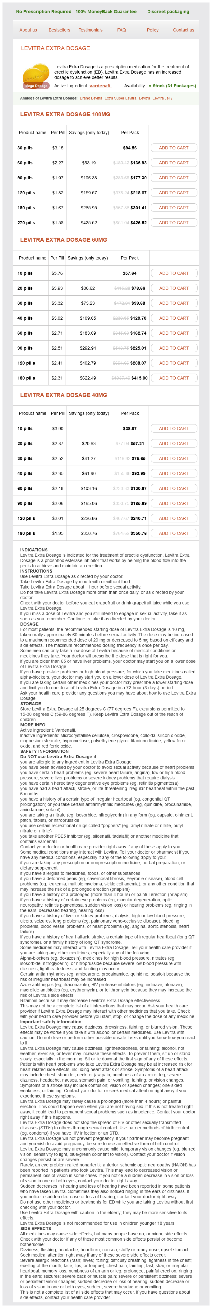
Levitra Extra Dosage Dosage and Price
Levitra Extra Dosage 100mg
- 30 pills - $94.56
- 60 pills - $135.93
- 90 pills - $177.30
- 120 pills - $218.67
- 180 pills - $301.41
- 270 pills - $425.52
Levitra Extra Dosage 60mg
- 10 pills - $57.64
- 20 pills - $78.66
- 30 pills - $99.68
- 40 pills - $120.70
- 60 pills - $162.74
- 90 pills - $225.81
- 120 pills - $288.87
- 180 pills - $415.00
Levitra Extra Dosage 40mg
- 10 pills - $38.97
- 20 pills - $57.31
- 30 pills - $75.65
- 40 pills - $93.99
- 60 pills - $130.67
- 90 pills - $185.69
- 120 pills - $240.71
- 180 pills - $350.76
The stomach starts emptying its contents within a few minutes of their reaching it vi erectile dysfunction treatment nj purchase generic levitra extra dosage on line. The stomach is higher in broad and stocky type in individual than in the slender type. Incisura angularis: At the junction of the body with the pyloric antrum the lesser curvature increases abruptly to form this angular notch. Lesser curvature: It runs almost vertically downwards to incisura angularis when it passes upwards and to the right, forming right border of stomach. Pyloric antrum: It is wide part of the pyloric region narrowing to pyloric canal, the terminal; part of which is surrounded by the pyloric sphincter. Pyloric canal: It appears as a column of barium, with parallel walls, about 2 to 3 mm wide and 5 to 8 mm long, joining the pyloric antrum. Its walls are smooth in outline and owing to the protrusion of the pyloric end in to the lumen of this part of duodenum b. Second part: It gives a floccular shadow because the barium emulsion is broken up in to all portions. Proximal part of small intestine the barium shadow remaining broken up and shows feather like appearance ii. Distal part of ileum forms a homologous shadow, coils are seen lying on the pelvis, last few inches are narrower than the rest. Cecum and the ascending colon: Sacculations known as haustrations, are present in proximal part of the colon but may not be evident in the distal part if the pressure is high iii. Owing to the acute angular curvature in the regions of the colic flexures and the pelvic colon b. Biliary Tract the method of visualization of the gall bladder is known as cholecystography. Oral cholecystography-telepaque (iopanoic acid) containing 66 percent iodine by weight is the oral preparation of choice. Intravenous cholecystography-it may be used for visualization of the gall bladder if diarrhea, pyloric obstruction or any other factors interferes with absorption of the orally administered contrast medium. It is usually seen in the angle between the twelfth rib and the upper lumbar vertebrae b. The density of the shadow is subject to considerable variation in normal individuals c. When the gall bladder is not visualized it may mean any one or more of the followings possibilities: i. Markedly impair liver function so that bile formation is impeded and the dye is not excreted ii. Obstructive disease of extrahepatic bile ducts so that the dye does not reach the gall bladder iii. Diseased gall bladder (chronic cholecystography) results in nonconcentration of the dye v. Nonfunction and occlusion of the gallbladder is demonstrated by the fact that the dye, although present in the bowel, is not seen in the gallbladder. To outlines the calyces, ureter, bladder, contain organic compounds containing iodine in their molecule, have to be introduced either intravenously or through a catheter to make the urinary tract radiopaque such as X-ray in which the urinary tract is visualized by a radiopaque medium is known as pyelogram. Generally the passage of drug through the kidney is too fast to reach a useful concentration b. Run downwards close to the tips of the transverse processes of the lumbar vertebrae and in front of the sacroiliac joints. In the pelvis they cast a shadow 1 cm on the medial side of the pelvic brim and across the tip of the ischial spine from where they run medially to join the bladder shadow. Ascending Pyelogram Radiographic appearance the following structures can easily be visualized: i. Uterine cavity: Appears as passing from the upper angles of the uterus and taking a tortuous course laterally ii. Paritoneal spill of the contrast median is a sign of patency of the fallopian tube. This is resorted to assess the configuration of the maternal pelvis as well as the age of the fetus ii. Acromioclavicular joint: As a gap between the clavicle and the acromion process of scapula ii. Acromion process: Lying partly behind the head of the humerus and superimposed on it iii. Medial portion is on the level with the junction of the middle and lower onethirds of the glenoid cavity b. Coronoid tubercle: As a bony prominence on the inferior surface of the clavicle near the outer third Female Genital Tract Hysterosalpingography i. This is particularly useful in cases of the sterility and to prove or disprove the patency of the uterine tube ii. Coronoid process: As a more or less circular shadow below the lateral one-third of the clavicle vii. Greater tubercle of humerus: As the most lateral bony point in the shoulder region ix. Inferior angle of scapula: As a partly superimposed on the lung field, at the level of the 7th rib or 7th intercostals space xi. Superior angle of scapula: Projects upwards in the angle between the clavicle and the first rib. As a translucent broad line passing across the ulna between the trochlea and coronoid process b. Lateral epicondyle of humerus: Seenasflutter appearance as compared to the medial epicondyle iv. Olecranon process of ulna: Superimposed on the shadow of the humerus and its proximal limit can be reorganized below the shadow, cast by the coronoid and olecranon fossae vi.
© 2025 Adrive Pharma, All Rights Reserved..