General Information about Isordil
If you would possibly be experiencing symptoms of angina or have been recognized with coronary heart disease, discuss to your doctor about whether or not Isordil could also be an acceptable therapy option for you. By selling rest and dilation of blood vessels, Isordil can help to enhance blood circulate to the heart and reduce the chance of angina attacks. With correct use and monitoring, this medication can be a helpful device in managing and preventing angina episodes.
It is crucial to take Isordil exactly as prescribed by a health care provider. Missing doses or stopping the medicine abruptly can result in a rise in the frequency and severity of angina episodes. It is important to also be aware of any other drugs or supplements that will interact with Isordil, as this will influence its effectiveness and enhance the risk of unwanted facet effects.
Angina is usually described as a feeling of stress or tightness within the chest, together with discomfort or pain. It is a common symptom of coronary heart illness, which occurs when there is a buildup of plaque within the arteries that offer blood to the heart. This plaque can restrict blood flow, leading to decreased oxygen and vitamins reaching the guts. As a outcome, angina can happen during bodily actions, similar to train and even sexual activity, when the guts is working more durable and requires more oxygen.
In addition to its use in preventing angina, Isordil can be used in the remedy of acute angina attacks. The medicine is out there in numerous varieties, together with tablets, sprays, and patches. The pill form is often taken by mouth and is absorbed shortly into the bloodstream, providing relief from angina symptoms inside minutes. The spray type is applied beneath the tongue and likewise works rapidly to relieve symptoms. The patch type is applied to the skin and slowly releases the medication over a interval of 24 hours.
Isordil is generally well-tolerated, but like all treatment, it could cause unwanted effects in some people. These can embrace headaches, dizziness, flushing, and nausea. Rarely, extra critical side effects similar to low blood strain, fast heart price, and fainting might happen. It is essential to discuss any potential unwanted effects with a doctor and to hunt medical consideration if any unusual symptoms occur.
Isordil is usually prescribed as a safety measure for angina, to be taken before engaging in physical actions that may set off an episode. It works by dilating the blood vessels, allowing for elevated blood circulate to the heart and lowering the workload on the center muscle. This might help to stop angina assaults and improve the overall functioning of the heart.
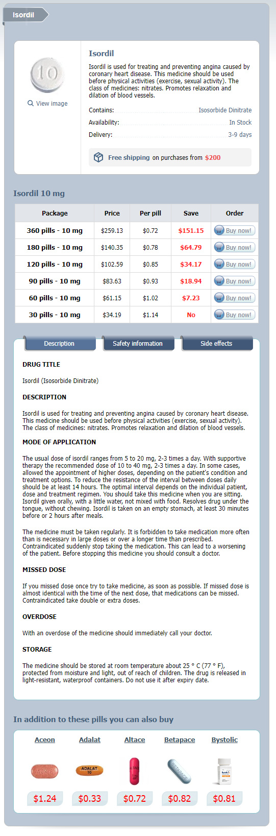
Isordil, also known as isosorbide dinitrate, is a medication that is primarily used for treating and stopping episodes of angina. Angina is a situation that occurs when there's a decreased blood move to the center, sometimes due to blockages in the coronary arteries. Isordil belongs to a class of medicines called nitrates, which work by promoting relaxation and dilation of blood vessels within the body. This allows for improved blood circulate and may help to alleviate the signs of angina.
In the classical pathway treatment centers for alcoholism buy generic isordil online, components are activated in the following order: C1, C4, C2, C3, C5, C6, C7, C8 and C9. The classical pathway is activated when C1q binds to IgM or IgG antibodies or C-reactive protein bound to microbe surfaces, or when C1q binds directly to molecules on bacterial surfaces such as lipoteichoic acid and phosphocholine. The other two pathways are termed innate pathways because they do not require antibody for activation. The alternative pathway is activated by the spontaneous hydrolysis of C3, which combines with two proteins, B and D. Serine proteases cleave components C2C5 and factor B into two fragments, for example, component C2 into C2a and C2b or component B into Ba and Bb. With the exception of C2, where the binding fragment is C2a, b fragments bind to the microbe surface and a fragments are soluble mediators of inflammation. Although the three pathways differ in the way that they are activated, they converge into a single effector pathway at the stage of formation of a C3 convertase. The function of the C3 convertase is to generate a large amount of C3b, which is covalently bound to the microbe. The addition of C3b to C3 convertase generates a C5 convertase that cleaves C5 into C5a and C5b. C3a and C5a fragments bind to receptors on endothelial cells, mast cells and macrophages, and C5a fragments also bind to receptors on neutrophils and initiate an inflammatory response. C8 initiates the polymerisation of between 10 and 16 molecules of C9 that create a pore in the cell membrane. Microbes opsonised by C3b are efficiently captured and destroyed by phagocytes bearing C3b receptors. C3b receptors on erythrocytes allow pathogens and pathogen molecules to be cleared from the circulation and degraded in the spleen and liver. Whereas the recruitment of soluble regulators by capturing host proteins is a common strategy to impair downstream complement actions, certain viruses also produce structural mimics of these regulators. Direct inhibition of C3, the C3 and C5 convertases, C5 or the C5a receptor (C5aR) is a prominent strategy of Staphylococcus aureus. Finally, a set of different microbial proteases can degrade many of the crucial components of the complement system. Increased and decreased activity is represented by thick and thin arrows, respectively. The complement system can target host cells as well as pathogens, so it is necessary that the cascade be tightly regulated. The complement cascade is such a powerful component of both innate and adaptive immunity that it is not surprising that pathogenic microorganisms have invested extensively in mechanisms that subvert it. OmpA outer membrane protein A StcE secreted protease of C1 esterase inhibitor TraT TraT outer membrane protein Fusobacterium spp. Bac -protein Fba fibronectin-binding protein Hicb factor H-binding inhibitor of complement IdeS IgG-degrading Enzyme of S. Complement-degrading proteases are utilised by bacterial pathogens such as Staphylococcus aureus (C3b-staphylokinase), Streptococcus pyogenes (C5a peptidase), S. Microbial envelope/wall components that inhibit complement the capsules of bacteria and fungi inhibit the deposition of complement components on their surfaces and/or inactivate them. In addition, capsules block access of antibody and C3b to the cell wall located beneath the capsule. For example, the capsule of Streptococcus pneumoniae impairs deposition of C3b and decreases conversion of C3b to iC3b. The capsules of Streptococcus agalactiae and Neisseria meningitidis contain sialic acid that prevents alternative pathway complement activity by creating a non-activating surface and by binding factor H, a plasma complement regulator. The transmembrane protein gC1 of herpes simplex virus type 1 and the transmembrane protein gC2 of herpes simplex virus type 2 bind C3b and increase the rate of breakdown of the alternative pathway C3 convertase. This observation has lead to the view that the opsonic and inflammatory components of complement may be more important in anti-pathogen immunity than pathogen lysis. For example, Trypanosoma bruci blebs off pieces of its membrane carrying away antibodies and complement components and T. Several bacteria liberate their capsule so that complement and antibodies bind capsule in the fluid phase rather than on the surface of the organism. Eosinophils and basophils are also phagocytic, but neutrophils and macrophages are the foremost cells involved in pathogen uptake and killing. In humans, neutrophils comprise about one-half to two-thirds of all blood leucocytes, and their number can increase 10-fold during infection. Macrophages begin their life either as progenitor cells that seed tissues during embryogenesis and self-renew throughout life or as monocytes. This is accomplished by recognition of chemoattractant gradients created by interaction of the pathogen with molecules of the innate immune system such as the complement components C3a, C4a and C5a that have been described above and by components of the pathogens themselves that are detected by phagocyte cell membrane receptors. The phagocyte may extend pseudopods that wrap around the pathogen, or the phagocyte membrane may invaginate to form a cup-shaped depression into which the pathogen sinks. In either case, the phagocyte cell membrane flows over the pathogen and seals it in a vacuole termed a phagosome. Phagosomes move towards the centrosome and, during their passage, their interior becomes increasingly acidic, reaching a terminal pH of about 4. In addition to lysosomes, neutrophils contain several types of granules that contain acidic hydrolases and antimicrobial proteins that fuse with phagosomes. Secretory vesicles/recycling endosomes contain cell membrane receptors that become incorporated in the formation of the phagosome and are recycled back to cell membrane of the phagocyte.
Because endurance and resistance exercise produces different results medicine 9 minutes isordil 10mg buy low price, an optimal exercise program should include both types of training. Myosin and actin myofilaments form cross bridges, and the actin pulls the myosin myofilament toward the center of the sarcomere. After forming cross bridges with the actin myofilament, the myosin myofilament propels the actin myofilament toward the center of the sarcomere. The sarcomere shortens, pulling the actin and myosin myofilaments toward the center, which pulls the Z-discs closer together. A continuous state of partial muscle contraction in which muscles are at their optimal resting length is called: a. The prefix bi- in a muscle name, such as in biceps brachii, refers to the fact that the muscle: a. During the process of muscle contraction, the sarcoplasmic reticulum is stimulated to release which substance Which muscle is often called the "praying muscle" because of its role in flexing the head Name the two types of cells that make up the nervous system and describe the function of each. Explain the process of impulse conduction in both myelinated and unmyelinated nerve fibers. List the 12 cranial nerves, using name and number and identify the functions of each. Identify the differences in structure and function between the sympathetic and parasympathetic divisions of the autonomic nervous system. To remain in balance (homeostasis), the various organ systems of the body must work together. Even an act as simple as eating lunch requires input from multiple body systems, including the endocrine system (which senses a drop in blood glucose levels and triggers the sensation of hunger), the muscular system (which allows you to chew your food), and the digestive system (which processes the food and eliminates the waste). The nervous system coordinates these systems so each knows exactly what to do and when to do it. The nervous system-consisting of the brain, spinal cord, and nerves-constantly receives signals about changes within the body as well the external environment. It then processes the information, decides what action needs to occur, and sends electrical and chemical signals to the cells, telling them how to respond. The nervous system also powers our ability to learn, feel, create, and experience emotion. Overview of the Nervous System the body has two organ systems dedicated to coordinating the activities of the trillions of cells making up the human form. One of those systems-the endocrine system-employs chemical messengers called hormones to communicate with cells. In contrast, the nervous system uses electrical signals to transmit messages at lightning speed. The nervous system has three essential roles: Sensing 1 the nervous system uses sense organs and nerve endings to detect changes both inside and outside the body. Integrating 2 3 the nervous system processes the information received, relates it to past experiences, and determines what response is appropriate. The nervous system issues commands to muscles and glands to initiate changes based on its information. Ganglia the peripheral nervous system consists of the vast network of nerves throughout the body. In brief, the peripheral nervous system consists of everything outside of the brain and spinal cord. Neurons are the excitable, impulse-conducting cells that perform the work of the nervous system, whereas neuroglia protect the neurons. Underscoring the importance of neuroglia is the fact that the nervous system contains about 50 glial cells for each neuron. Schwann cells are found in the peripheral nervous system; all the rest reside in the central nervous system. This arrangement allows the astrocyte to funnel glucose from the bloodstream to the neuron for nourishment. However, it also prevents most medications from reaching brain tissue, making treating disorders of the brain challenging. Although this allows them to replace worn-out or damaged cells, it also makes them susceptible to tumor formation. There are three classes of neurons: sensory (afferent) neurons, interneurons, and motor (efferent) neurons. Each neuron type fulfills one of the three general functions of the nervous system. Besides receiving, processing, and storing information, the connections made by these neurons make each of us unique in how we think, feel, and act. Dendrite Multipolar neurons Multipolar neurons have one axon and multiple dendrites. This is the most common type of neuron and includes most neurons of the brain and spinal cord. Axon branch Dendrite Bipolar neurons Bipolar neurons have two processes: an axon and a dendrite with the cell body in between the two processes.
Isordil Dosage and Price
Isordil 10 mg
- 360 pills - $259.13
- 180 pills - $140.35
- 120 pills - $102.59
- 90 pills - $83.63
- 60 pills - $61.15
- 30 pills - $34.19
Bioengineering and regeneration of the endocrine pancreas Pancreas development 173 2° transition B treatment 5th toe fracture discount isordil 10mg buy on line. Spatiotemporally expressed transcription factors regulate pancreatic cell fates within the trunk and tip domains. On the other hand, trunk cells are demarcated by high levels of Oc1, Sox9, Hnf1, and Nkx6. The cells that line the cylindrical tube-like luminal "trunk" domain are bipotent in nature and give rise to either endocrine or ductal cells. Exocrine versus endocrine development during secondary transition the secondary transition (occurring at approximately e13. The spatiotemporal manner by which exocrine and endocrine differentiation occurs is a crucial component to a thorough understanding of cell lineage allocations in the pancreas. Cell lineage determinations in the developing pancreas are most often described by their unique transcriptional programs. Below we cover the different transcription factors and the downstream targets that are involved in pancreas cell fate allocations. Exocrine cell fate allocation Acinar and ductal cells make up the exocrine portion of the pancreas. Acinar cells are considered the functional unit of the exocrine pancreas where they synthesize, store, and secrete digestive enzymes into the ductal epithelial network, ultimately leading to the duodenum. Although the exocrine compartment of the pancreas holds significant physiological and pathophysiological relevance, this chapter focuses on the developmental processes of the endocrine portion of the pancreas. Endocrine cell fate allocation Endocrine lineage allocation is primarily regulated by the pro-endocrine transcription factor Neurog3, which is induced by combined activity of the Oc1 and Pdx1 transcription factors. Neurog3 expression is initially observed in scattered cells within the prepancreatic epithelium at e9. Transgenic overexpression of Neurog3 throughout the mouse pancreatic epithelium results in an increase in endocrine specification, but generates only increased numbers of -cells B. Embryonic development of the endocrine pancreas (the first hormone-expressing cells to differentiate). The earliest Neurog3-expressing progenitors favor -cell commitment whereas - and -cells arise from progenitors demonstrating Neurog3 expression at later timepoints. Through an asymmetric cell division, Neurog+ cells generate an apical daughter cell that maintains a connection with the epithelial layer and a basal daughter cell that does not; apical cells conserve their progenitor-like capabilities while basal cells differentiate into endocrine cells. Delamination results from Snail2 activation (within Neurog3+ cells) leading to inhibition of E-cadherin expression, thus allowing endocrine progenitors to migrate radially into the parenchyma from the epithelial trunk domain in an epithelial-tomesenchymal transition. Endocrine cell subtype specification: - and -cells Although endocrine progenitors expressing Neurog3 exhibit reduced Pdx1 expression, cells destined for the -cell lineage upregulate Pdx1. Many studies have demonstrated a clear, essential role for Pdx1 in -cell specification and differentiation. Pdx1 activity simultaneously promotes -cell differentiation while repressing the -cell fate. Transgenic overexpression of Pdx1 in glucagon-expressing cells postnatally results in -to- cell conversion, while hypomorphic Pdx1 alleles result in increased numbers of -cells and decreases in -cells, leading to impaired glucose tolerance. Expression of Arx in the pancreatic epithelium during development and in adult -cells increases the -to-/-cell ratio. In fact, Pax4 and Arx have been shown to directly inhibit the expression of the other through direct promoter binding, suggesting they both directly and indirectly reciprocally repress their respective endocrine cell fates. In contrast to other broadly expressed transcription factors, MafA and MafB are only expressed in hormone-positive cells. The MafA and MafB expression patterns in mouse insulin+ cells are dynamic in nature with MafB being expressed in immature insulin there should be no spaces between the word before the + sign and the + sign. In mice, there is a developmental transition from MafB to MafA expression in -cells. In contrast, in humans MafB expression is not silenced in -cells but remains elevated throughout adulthood concomitant with MafA. Moreover, initiation of MafA expression occurs significantly earlier in mice (secondary transition) whereas onset of MafA expression in humans is roughly around age 11. Somatostatin-secreting -cells play physiologically important roles in islet function, inhibiting both insulin and B. Bioengineering and regeneration of the endocrine pancreas Postnatal islet development and function 177 glucagon secretion, but somatostatin itself does not appear to have any role in normal pancreas development. Glucose responsiveness, a characteristic trait of postnatal islet maturation, is regulated by an intricate network of genes that are expressed after birth. Historically, mouse islets were considered structurally and functionally mature upon expression of MafA and Glut2 concomitant with a loss in MafB. However, recent studies have demonstrated that urocortin 373, 74 and synaptotagmin 4 are also molecular markers of -cell maturation. Species-to-species distinctions in islet composition and morphogenesis exist in mammals15, 76, 77 though the precise underlying mechanisms governing these differences have not been fully characterized. Pancreatic endocrine cells initially assemble into a mantle-core islet morphology during the early postnatal period. In adults, mouse, rat, and pig islets have Endocrine development during late gestation There are several factors involved in pancreas development that have their main effect on endocrine differentiation during late gestation. For example, Foxa2 is present during the very early stages of pancreatogenesis but does not appear to affect development until late gestation, when it represses terminal differentiation of -cells.
© 2025 Adrive Pharma, All Rights Reserved..