General Information about Haldol
One significant danger related to Haldol is its potential to trigger a uncommon however severe aspect impact often recognized as neuroleptic malignant syndrome (NMS). NMS is a severe neurological situation that may happen in individuals taking antipsychotic medication, including Haldol. It is characterised by symptoms such as high fever, muscle rigidity, altered psychological standing, and unstable blood pressure. If left untreated, NMS may be life-threatening, which is why it's crucial for individuals taking Haldol to be carefully monitored for any signs of this situation.
Haldol can also enhance the risk of developing a movement dysfunction often recognized as tardive dyskinesia (TD). TD is characterised by involuntary movements of the face, jaw, and tongue, and can be irreversible in some circumstances. The risk of developing this situation is considerably larger in people taking Haldol for extended durations, particularly in those over the age of sixty five. Therefore, regular monitoring and dosage changes are needed to reduce the chance of TD.
Schizophrenia is a psychological dysfunction characterised by a variety of symptoms, together with hallucinations, delusions, disordered pondering and behavior, and a lack of motivation. It impacts roughly 1% of the worldwide population and is understood to have a major influence on the day-to-day functioning of individuals, as nicely as their relationships and general quality of life. The actual reason for schizophrenia isn't absolutely understood, but research has proven that it could be due to a combination of genetic, environmental, and neurochemical components.
In latest years, there has been a development in the course of prescribing newer, second-generation antipsychotics over typical antipsychotics like Haldol. This is as a outcome of of their lower danger of side effects and potential for better outcomes. However, Haldol stays a priceless and widely used treatment, particularly in instances the place different therapy choices have been unsuccessful or are not affordable. It is commonly used as a first-line remedy for schizophrenia and is still most well-liked by some healthcare providers because of its efficacy and decrease cost.
Haldol can additionally be used to treat individuals with Tourette's syndrome, a neurological situation characterized by repetitive, involuntary movements and vocalizations generally known as tics. While the exact cause of Tourette's is not fully understood, it's believed to be due to an abnormality in the brain's neurotransmitter systems, together with dopamine. Similar to its results on schizophrenia, Haldol works by blocking dopamine signaling, which might reduce the frequency and severity of tics in people with Tourette's.
Haldol belongs to a class of medication generally recognized as typical or first-generation antipsychotics. These medicines work by blocking dopamine, a neurotransmitter in the brain that is involved in regulating mood, habits, and cognition. Excess dopamine exercise is assumed to contribute to the signs of schizophrenia, and Haldol's capability to inhibit it helps to minimize back these symptoms. It is important to note that Haldol isn't a cure for schizophrenia, but it could successfully manage signs and improve the standard of life for these dwelling with the disorder.
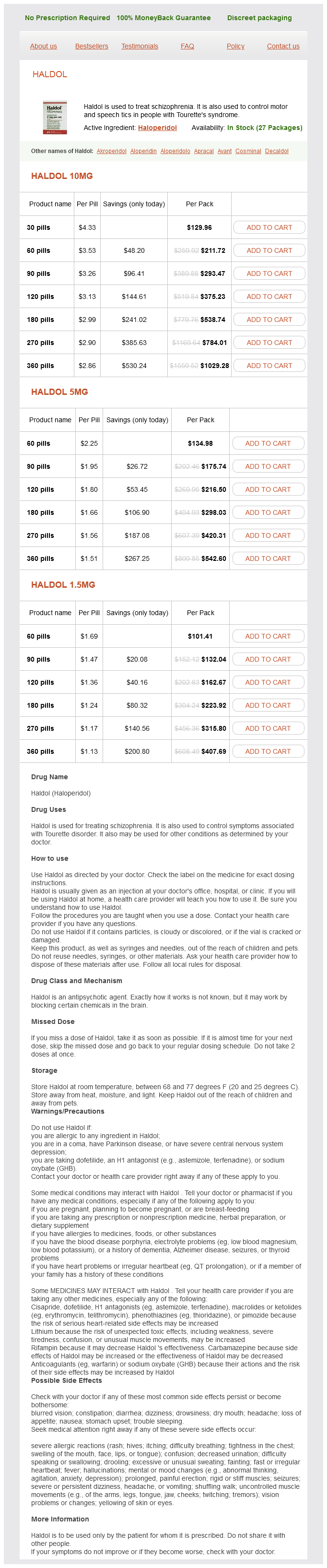
Haldol, also known by its generic name haloperidol, is a drugs used to treat a quantity of mental and neurological disorders, most notably schizophrenia. It can be efficient in managing motor and speech tics in people with Tourette's syndrome. Haldol is a powerful antipsychotic drug that has been available on the market for over 50 years and remains a commonly prescribed medicine for those suffering from these conditions.
Despite its effectiveness in treating these situations, Haldol is not without its potential unwanted side effects. The most common unwanted side effects embrace drowsiness, dizziness, nausea, constipation, and dry mouth. Some people may expertise more serious unwanted facet effects similar to muscle stiffness, tremors, restlessness, and involuntary movements of the face and body. These unwanted effects could be managed with appropriate dosing and close monitoring by a healthcare skilled.
In conclusion, Haldol is a potent treatment that has been used for many years to manage symptoms of schizophrenia and motor and speech tics in individuals with Tourette's syndrome. While it is highly effective, it could be very important fastidiously monitor its utilization and potential unwanted aspect effects. Ultimately, healthcare providers must weigh the potential dangers and advantages of Haldol for each particular person affected person to discover out one of the best course of therapy. With correct monitoring and accountable utilization, Haldol remains an invaluable treatment within the treatment of these complicated and difficult conditions.
Median nerve formed by its two roots medicine of the prophet cheap haldol 5 mg fast delivery, which unite anterior to the axillary artery, it runs down the arm anterior to the brachial artery, overlapped by the bicipital aponeurosis, into the cubital fossa lying medial to the artery. Ulnar nerve largest branch of the medial cord, it runs medial to the axillary artery and just posterior to the medial cutaneous nerve of the forearm. Halfway down the arm the ulnar nerve passes into the posterior compartment to continue its downwards course superficial to triceps; at the elbow it lies posterior to the medial epicondyle of the humerus, where it is palpable and most vulnerable to damage. Medial cutaneous nerve of the forearm almost as large as the ulnar nerve, but lying anterior to it (as might be expected since it is heading for skin) and not to be confused with it. Radial nerve largest nerve of the brachial plexus, from the posterior cord, posterior to the axillary artery; anterior to the wide tendon of latissimus dorsi on the lower posterior axillary wall. It is the nerve of the extensor muscles in the arm and forearm (including brachioradialis). Radial nerve injury from fracture of the humerus does not usually paralyse triceps because the branches that supply it arise high in the axilla above the level of injury. It curls around posterior to the humerus in the radial groove, between the medial and lateral heads of triceps, to emerge laterally deep to brachioradialis to innervate it and all the extensors in the forearm. It divides into a relatively unimportant superficial cutaneous branch and the highly important deep radial nerve, which carries the motor supply to all the forearm extensor muscles. The deep radial nerve runs between the two heads of the supinator and emerges distally as the posterior interosseous nerve. Remember, therefore, that the radial nerve, which comes from the posterior cord of the brachial plexus, is the nerve that supplies the muscles of the posterior aspect of the arm and forearm. Axillary nerve large nerve arising high up from the posterior cord, it runs downwards and laterally to disappear posteriorly between the tendons of subscapularis and teres major and the humerus, to innervate the deltoid (and teres minor) and, clinically important, a small overlying patch of skin inferior to the acromion. They are divided into groups (anterior or pectoral group, posterior and lateral), all draining to a central group, which in turn drain to an apical group in the axillary apex. The axillary lymph nodes are commonly invaded by cancerous spread (metastases) from the breast one of the commonest sites for cancer in females. Coracobrachialis from the coracoid process of the scapula (with the short head of biceps) passing halfway down the medial side of the humerus. Very weak flexor of the shoulder joint and notable because the musculocutaneous nerve runs through and innervates it a useful identifying feature. Triceps extensor of the elbow (with the long head also weakly extending the shoulder), the largest muscle on the posterior of the arm, with heads of origin from the scapula inferior to the glenoid cavity (long head), the upper part of the posterior of the humerus (lateral head) and the rest of the posterior of the humerus (medial head). Anconeus a very small triangular muscle from the posterior surface of the lateral humeral epicondyle passing distally to the posterior surface of the ulna. In the upper (proximal) part of the arm the brachial pulse can be felt by pressing laterally, not backwards, because at this level the artery lies medial to the humerus, not in front of it. Apart from receiving lymph from the upper limb, they are of supreme clinical importance because most of the lymphatic drainage from the breast passes to these nodes. Biceps the prominent muscle on the anterior of the arm, with a long head originating from the supraglenoid tubercle within the shoulder joint, and a short head arising from the coracoid process with coracobrachialis. At the elbow its tendon is attached to the posterior of the tuberosity of the radius. It is not only a flexor of the elbow joint (and a weak flexor of the shoulder), but also (with the elbow flexed and forearm pronated) the most powerful supinator of the forearm (p. There is a thin expansion (bicipital aponeurosis) of the tendon, which passes superficially and medially to lie between the antecubital veins, commonly used for venepuncture, and the deeper located brachial artery and median nerve. Brachialis deep to biceps, from the anterior of the distal humerus to the anterior of the coronoid process and tuberosity of the ulna. It is a powerful flexor of the elbow joint innervated by the musculocutaneous nerve. To understand these, flex your elbow to a right angle and look at the palm of the hand (supine position), then turn the hand over so that you are looking at the dorsum of the hand (placing it in the prone position). This is the movement of pronation, where the lower end of the radius (the lateral bone of the forearm) rotates round the lower end of the ulna (the medial bone of the forearm), carrying the hand with it. Now turn the hand over so that you are looking at the palm (supine) again; this is the movement of supination. For many common actions, like holding a glass, the forearm and hand are used in the mid-prone position, midway between full pronation and full supination. The ligaments of the radioulnar joints and the fibrous interosseous membrane stretching between the radius and ulna keep the two bones together during these movements. The medial epicondyle gives origin to several flexor muscles and forms the common flexor tendon; similarly, the common extensor tendon attaches to the lateral epicondyle. Any of these bony prominences are easily hit against objects and a resultant fracture of the more prominent medial epicondyle can damage the ulnar nerve, which lies in close contact. Elbow, forearm and hand the power and the range of upper limb activity are enormous, extending from the relatively crude movements of wielding a hammer to the most delicate brush strokes of the artist or the steady manipulations of the neurosurgeon. The coordination of motor and sensory activities in the hand is matched only by those of the eye. The twisting movements of the forearm that turn over the hand and the unique rotatory movement at the base of the thumb, allowing it to be carried towards the palm of the hand to give a firm grip, have given a degree of manual dexterity that has contributed to With the elbow straight (extended), the head of the radius can be felt on the posterior aspect of the elbow (at the bottom of a small depression lateral to the olecranon), K30266 Book. At the sides of the wrist, the styloid process of the radius extends 1 cm distal to the styloid process of the ulna. It contains, from lateral to medial, the tendon of biceps, the brachial artery and the median nerve. The radial nerve is deep to brachioradialis on the lateral side and so is not visible unless the muscle is displaced laterally, where the nerve can be seen dividing into its superficial (cutaneous) and deep (posterior interosseous) branches. Pronator teres arising proximally from the common flexor origin, the muscle crosses the forearm obliquely to be attached distally halfway down the lateral side of the radius. It has a small deep head from the coronoid process of ulna, and the median nerve, by which it is innervated, passes distally between the two heads. Brachioradialis from the lateral side of the humerus proximal to the lateral epicondyle, the muscle runs distally to the lower end of the radius just proximal to the styloid process.
The junctions between the shafts and epiphyses of developing bones are also a type of cartilaginous joint medicine vials 5 mg haldol purchase visa, although they disappear as growth ceases. The bone ends are covered by cartilage and surrounded by a fibrous capsule that encloses a joint cavity. The capsule is reinforced by ligaments on the outside and sometimes has other ligaments inside. The inside of the capsule is lined by synovial membrane, which secretes a minute amount of synovial fluid (the knee joint, the largest, has only 0. Synovial joints allow varying degrees of movement and, depending on the shape of the articulating surfaces, can be classified into various types: balland-socket (hip, shoulder), hinge (elbow, interphalangeal joints of fingers and toes), condylar (modified hinge, as at the knee and temporomandibular, or jaw, joint), ellipsoid (modified ball-and-socket, as at the wrist), saddle (saddle-shaped surfaces, as at the base of the thumb) and plane (rather flat surfaces, as between some wrist and foot bones). The details of individual joints are considered in the chapters for the appropriate regions. Cranium strictly means the skull without the mandible, but is often used to mean the upper part of the skull that encloses the brain; it is made up of paired parietal and temporal bones and of single occipital, sphenoid, ethmoid and frontal bones. External features are considered below and internal features in Chapter 3 (Head, neck and vertebral column, p. Underlying it on the inside is a branch of the middle meningeal artery, liable to be K30266 Book. Facial skeleton the front (anterior) part of the skull, containing the orbital and nasal cavities. The principal bones are the single mandible (lower jaw with lower teeth) and paired zygomatic bones and maxillae (forming the upper jaw with upper teeth), with the frontal bone forming the forehead. The margins of each orbit are formed by the frontal and zygomatic bones and maxilla. Posterior nasal apertures (choanae) above the back of the hard palate, opening into the nasal part of the pharynx. Mandibular fossa in the temporal bone, forming the temporomandibular joint (jaw joint) with the head of the mandible. Occipital condyles on either side of the foramen magnum, forming atlanto-occipital joints with C1 vertebra (atlas). It consists of a central body and a greater horn on each side, with a much smaller lesser horn projecting up from the junction between the body and greater horn. Various muscles and ligaments are attached to it, but it is unique in that it makes no joint with any other bone. Each vertebra typically consists of a body anteriorly, with a vertebral (neural) arch posterior to the body. The arch is made up of a pedicle (attached to the body) on each side and a lamina posteriorly; two laminae unite in the midline to form the spinous process. When articulated, the gap between the pedicles of adjacent vertebrae, bounded posteriorly by the zygapophyseal (commonly called facet) joints and anteriorly by the intervertebral disc, forms the intervertebral foramen, the important opening through which each spinal nerve emerges (p. The first cervical vertebra is also called the atlas (unique in that is has no body), which makes joints on each side with the skull above (atlanto-occipital joints) and with the second cervical vertebra, the axis, below (lateral atlanto-axial joints). The remaining cervical vertebrae and the thoracic and lumbar vertebrae are united by various ligaments, in particular the anterior and posterior longitudinal ligaments (each of which is a long continuous band on the anterior and posterior surfaces, respectively, of the vertebral bodies) and small joints between the adjacent articular processes (zygapophyseal or facet joints). Each consists of outer concentric rings of fibrocartilage that form the annulus fibrosus, with a more centrally located gelatinous mass, the nucleus pulposus. The highest disc is the one between the C2 (axis) and the C3 vertebrae; the lowest (the one most commonly prolapsed) is between the L5 vertebra and S1 of the sacrum. It is joined above to the fifth lumbar vertebra by an intervertebral disc and ligaments and laterally to the hip bones through the sacroiliac joints to form the bony pelvis, and at its lower end it is joined with the coccyx (of four rudimentary coccygeal vertebrae) through the sacrococcygeal joint. Each rib has a head, which typically articulates with the bodies of two adjacent vertebrae, a neck, a tubercle (which articulates with the transverse process of its own vertebra) and a body or shaft of variable length that forms the curved chest wall. The first seven pairs of ribs (true ribs) are joined to the sternum by their costal cartilages. The next three pairs (false ribs) are joined by their cartilages to the cartilage above. The sternum consists of the manubrium (at the top cranial end), body and xiphoid process (at the lower caudal end). Together the ribs, costal cartilages and the 12 thoracic vertebrae form the skeleton of the thorax. The manubrium and body are not quite in a vertical line, but unite at a slight angle (the sternal angle of Louis) to each other, forming the cartilaginous manubriosternal joint. The important manubriosternal joint locates the articulation of the second costal cartilage, which is useful when clinically locating specific intercostal spaces. At the distal end there is a prominent medial epicondyle and a less obvious lateral epicondyle. Between the two are the smooth articular surfaces for the elbow joint: medially, the pulley-shaped trochlea (for the ulna) with a prominent medial lip; and laterally, the rounded capitulum (for the radius). Posteriorly at the distal end is the deep olecranon fossa, which accommodates the olecranon of the ulna when the elbow is extended. Radius lateral bone of the forearm: has a rounded proximal end, the radial head, which articulates with the capitulum of the humerus and a notch on the ulna. The shaft immediately distal to the head is the neck, distal to which on the medial side, is the radial tuberosity (for attachment of the biceps tendon). Distally, the radial shaft is expanded to articulate with the carpal bones to form part of the wrist joint, and it ends by forming the point-like styloid process. Ulna medial bone of the forearm, with the proximal end deeply depressed anteriorly, forming the trochlear notch (whose posterior boundary is the olecranon) for articulation with the trochlea of the humerus. The small rounded distal end comprises the head, with a styloid process on its medial side. The eight small carpal bones each have their own characteristic sizes and shapes, details of which need not be learned. The important point is to remember the order of the bones in the two rows of four from the lateral to the medial side: in the proximal row, the scaphoid, lunate, triquetral Upper limb bones Clavicle rather S-shaped, with a bulbous medial end for the sternoclavicular joint and a flattened lateral end for the acromioclavicular joint, and a groove on the under surface.
Haldol Dosage and Price
Haldol 10mg
- 30 pills - $129.96
- 60 pills - $211.72
- 90 pills - $293.47
- 120 pills - $375.23
- 180 pills - $538.74
- 270 pills - $784.01
- 360 pills - $1029.28
Haldol 5mg
- 60 pills - $134.98
- 90 pills - $175.74
- 120 pills - $216.50
- 180 pills - $298.03
- 270 pills - $420.31
- 360 pills - $542.60
Haldol 1.5mg
- 60 pills - $101.41
- 90 pills - $132.04
- 120 pills - $162.67
- 180 pills - $223.92
- 270 pills - $315.80
- 360 pills - $407.69
Antimicrobial therapy is necessary in patients with chronic diarrheal disease symptoms 0f heart attack discount 5 mg haldol with amex, wound infections, or systemic disease. Antibiotic therapy, although of secondary value, can reduce toxin production and clinical symptoms and decrease transmission by the more rapid elimination of the organism. A single dose of azithromycin is currently the drug of choice for children and adults because macrolide resistance is relatively uncommon. A single dose of doxycycline or ciprofloxacin in nonpregnant adults can be used as alternative therapy if demonstrated to be active in vitro; however, resistance to the tetracycline and fluoroquinolones is relatively common. The combination of minocycline or doxycycline with ceftriaxone or cefotaxime appears to be the most effective treatment. Although long-term carriage of 26 · Vibrio and Related Bacteria 277 Clinical Case 26. Leeches remove stagnant blood and stimulate oozing of blood into the skin graft for up to 48 hours after removal of the leech. This bleeding is mediated by an inhibitor of thrombin, hirudin (source of the genus name), which is present in the saliva of leeches. Aeromonas is present in the leech gut and produces proteolytic enzymes used by the leech to digest blood. One complication of using leeches is wound infections with Aeromonas, as illustrated by the patient described by Snower and associates (J Clin Microbiol 27:14211422, 1989). A 62-year-old woman had basal cell epitheliomas removed from her forehead, with the surgical site covered with skin grafts. The leeches were removed from a leech tank and applied to the wound for 1 hour on four separate occasions. Eleven days after the initial surgery, the graft appeared infected and was removed. Cultures of this graft, and leeches and water from the leech tank, were positive for Aeromonas. The patient was treated with parenteral antibiotics, and regrafting without the use of leeches was successful. A fluoroquinolone can be used initially for empirical therapy, but activity should be confirmed with in vitro susceptibility tests. Thus the long-term effectiveness of fluoroquinolones Case Study and Questions A 57-year-old man was hospitalized in New York with a 2-day history of severe watery diarrhea. The patient was dehydrated and suffering from an electrolyte imbalance (acidosis, hypokalemia). He made an uneventful recovery after fluid and electrolyte replacement was instituted to compensate for the losses resulting from the watery diarrhea. Which antibiotics are generally effective against Pseudomonas but not Stenotrophomonas, and against S. A 70-year-old man who had been admitted 7 days previously to the intensive care unit for acute shortness of breath and a temperature of 39° C developed a new productive cough and associated pleuritic chest pain. Examination of his chest revealed crackles at the bases of both lungs, with rhonchi present in both upper lobes; the chest radiograph indicated bilateral opacities consistent with bronchopneumonia. Sputum and blood cultures were performed, and 24 hours later the laboratory reported isolation of Pseudomonas aeruginosa. To complicate our understanding of these organisms, their taxonomic classification has undergone numerous changes in recent years. Despite the many genera, most clinically significant isolates are members of five genera: Pseudomonas, Burkholderia, Stenotrophomonas, Acinetobacter, and Moraxella (Table 27. Pseudomonas the genus Pseudomonas originally consisted of a large heterogeneous collection of nonfermentative bacteria that were grouped together because of their morphologic 278 similarity. In 1992, this genus was subdivided into a number of new genera (including Burkholderia and Stenotrophomonas); however, there are still more than 250 species in Pseudomonas. Members of the genus are found in soil, decaying organic matter, vegetation, and water. Unfortunately, they are also found throughout the hospital environment in moist reservoirs such as food, cut flowers, sinks, toilets, floor mops, respiratory therapy and dialysis equipment, and even disinfectant solutions. It is uncommon for carriage to persist in humans as part of the normal microbial flora, except in hospitalized patients and ambulatory, immunocompromised hosts. They are capable of using many organic compounds as sources of carbon and nitrogen, and some strains can even grow in distilled water by using trace nutrients. These organisms also possess many structural factors, enzymes, and toxins that enhance their virulence and render them resistant to most commonly used antibiotics. Indeed, it is surprising that they are not more common pathogens, considering their ubiquitous presence, ability to grow in virtually any environment, virulence properties, and resistance to many antibiotics. Additionally, expression of virulence traits is regulated by complex cell-density signaling (quorum sensing) systems that in turn are influenced by host factors such as the presence of serum and cytokines. The organisms utilize carbohydrates through aerobic respiration, with oxygen the terminal electron acceptor. Although described as obligate aerobes, they can grow anaerobically using nitrate or arginine as an alternate electron acceptor. The presence of cytochrome oxidase (detected in a rapid 5-minute test) in Pseudomonas species is used to differentiate them from the Enterobacteriaceae and Stenotrophomonas. Despite the diversity of virulence factors, most experts believe that multiple factors must work together for P. Adhesins As with many bacteria, adherence to host cells is critical for establishing infection.
© 2025 Adrive Pharma, All Rights Reserved..