General Information about Fluconazole
Fluconazole, generally known by its brand name Diflucan, is an antifungal antibiotic used to treat a variety of fungal infections. It is a powerful medicine that is ready to effectively fight candidiasis, a type of fungal infection attributable to the Candida species of yeast.
Another advantage of fluconazole is its convenient dosing regimen. As mentioned earlier, most yeast infections may be handled with a single dose. This means that patients wouldn't have to stick to a complicated medicine schedule, making it simpler to complete the complete therapy course and achieve a successful consequence.
Aside from genital yeast infections, fluconazole can also be used to treat other kinds of candidiasis, such as thrush (a yeast infection within the mouth), skin and nail infections, and esophagitis (an infection of the esophagus). It can additionally be effective against sure kinds of fungal pneumonia and meningitis. In some cases, it could also be used as a safety measure in patients with weakened immune systems, similar to these with HIV/AIDS, most cancers, or organ transplants.
Candidiasis can have an result on varied components of the physique, together with the mouth, skin, and genitals. When it occurs in the genital area, it's generally referred to as a yeast infection. This kind of infection impacts both men and women, although it's extra common in women. The symptoms of a genital yeast an infection can include itching, burning, and irritation in the affected area, in addition to thick, white discharge.
However, you will want to notice that fluconazole may not be efficient against all types of yeast infections. In specific, it will not be effective towards infections caused by different kinds of fungi. Additionally, it's not really helpful to be used in pregnant ladies as it could harm the creating fetus.
Fluconazole works by targeting the cell membrane of the fungus, disrupting its growth and stopping it from reproducing. This means that the infection is unable to unfold and eventually dies off, permitting the body to heal. It is normally prescribed as a single oral dose for uncomplicated yeast infections, but may require a longer course of therapy for extra severe or recurrent infections.
One of the major advantages of fluconazole is its excessive efficacy and security profile. It is usually well-tolerated, with few unwanted side effects reported. The most common side effects include nausea, diarrhea, and dizziness, that are usually gentle and momentary. In uncommon cases, critical allergic reactions could happen, and quick medical consideration should be sought if these signs arise.
In conclusion, fluconazole is a extremely efficient medicine for treating numerous types of candidiasis, together with genital yeast infections. Its high efficacy, safety profile, and handy dosing make it a popular choice amongst healthcare providers and sufferers alike. However, you will need to comply with your physician's instructions and report any potential unwanted facet effects to make sure a successful treatment outcome. If you are affected by any kind of fungal an infection, do not hesitate to seek the advice of along with your physician and ask if fluconazole may be an option for you.
Fluconazole may also interact with other medicines, so you will want to inform your healthcare supplier of another drugs you're taking to ensure there are no potential drug interactions.
Motor deficits that include incoordination and hypotonic muscles may also be seen anti fungal meds for dogs buy fluconazole us. These signs may be seen in persons with mental disability as well as in highly functioning individuals. The development of this disease has been attributed to genetics and environment factors. Studies have shown irregularities in several regions of the brain of autistics, for example, the cingulate gyrus, which are normally associated with timing of brain development. Autistics utilize different parts of the brain in performing social or nonsocial functions. As a result, the brain may tend to develop at a quicker pace subsequent to unusual neuronal growth that produces abnormal neuronal synaptic connectivity, impaired neuronal migration, and unbalanced interaction between neurons. Preliminary data indicate that genetic mutations, deletions, duplications, and/or impact of environmental factors (chemicals such as in heavy metals, bacteria or viral infections, smoking and pesticides) and teratogens on genes disrupt the normal early fetal brain development and growth and adversely affect the synaptic neuronal connectivity. Prevalence of autism appears significantly more commonly in individuals with 1q21. Indications that autistics show genetic alteration linked to chromosome 15 (15q13. Abnormal levels of serotonin in the brains of autistic children have been reported. Data have been compiled regarding the role of metabotropic glutamate receptors and growth hormones in this disease. It has been reported that the brains of autistics have a structurally distorted "mirror neuron system" circuit, which regulates understanding and modeling of their reactions toward the gestures, emotion, intention of others. A poor connectivity and imbalance exist in autistics between parts of the brain that mediate social Telencephalon 167 functions and those areas that regulate attention and goal-directed tasks. Thus, stimuli associated with audition, vision, language, and facial recognition are processed differently in autistics. There is also evidence that in autistics, the connection between the frontal cortex and the rest of the neocortex is weak and that this poor connectivity is brain hemispheric and predominantly confined to the association cortices (underconnectivity theory). The limitation of this theory is evident in the ability of autistics to execute some functions without deficits. Disturbances of social cognition are also explained on the basis of the empathizing systemizing theory, which revolves around the fact that autistics have difficulty in assessing gestures and activities of others (empathizing) while remaining capable oft systemically controlling events generated by the brain (self). This type of selectivity in systemic versus empathic processing is further expanded to "extreme male brain theory," in which the male brain can perform systemization but is unable to produce empathic response. There are other theories that discuss disturbances of nonsocial "executive" functions in autistics. Children under the age of 10 years, and rarely, adults, exhibit frequent seizures, loss of motor skills, aphasia, and hemiparesis that progressively worsens. Wada test is a test conducted before brain tumor resection or surgical procedure utilized in the treatment of epilepsy. It is utilized to establish cerebral dominance and to determine the cerebral hemisphere that performs speech and memory functions. In this procedure, sodium amobarbital is injected into each internal carotid artery, one at a time. Therefore, the language and memory centers are blocked on one side in order to test the same functions in the opposite hemispheres. A brief aphasia, which accompanies injection into the internal carotid artery, may determine the dominant hemisphere. This procedure can produce a variety of complications, including contralateral hemiplegia and sensory neglect as well as manifestations of disinhibition. The amygdala is a structure that maintains connections with the striatum, thalamus, cerebral cortex, and structures that constitute the limbic system (see Limbic System, Chapter 17). Both the caudate nucleus and putamen form the neostriatum, representing the afferent portion of the basal nuclei, whereas the lentiform nucleus refers to both the putamen and the globus pallidus. For further discussion of the basal nuclei, see Extrapyramidal Motor System, Chapter 21. Knowledge of anatomy of the cerebral vessels is of utmost clinical importance, as it is essential in performing and interpreting angiographic imaging, understanding the deficits associated with vascular accidents, and developing a treatment plan. The blood supply to the brain tissue is maintained by the carotid and vertebrobasilar arterial systems. There are specific features of the capillaries associated with these two arterial systems. For instance, these capillaries are composed of nonfenestrated continuous endothelial cells, connected by tight junctions, and encased by perivascular end feet of astrocytes, possessing large number of mitochondria and pinocytotic vesicles. Approximately 15% of cardiac output, equivalent to 750 mL per minute, is directed to the brain. The difference between the arterial and venous pressure may determine the cerebral blood flow. In addition, intrinsic factors such as the condition of the cerebral vessels, changes in the tension of carbon dioxide, and alteration in pH will also affect the blood flow to the brain. Due to the small size of frontal lobes in infants, the cerebral vessels are concentrated within the Sylvian triangle. Vascular disorders are occlusive in nature, in which the dysfunction is a sequel to ischemia or hemorrhage. Cerebral emboli may occur at any time, but it is especially common during daytime. They may originate from the pulmonary veins, cardiac valves or chambers, and plaques in the aortic arch or its branches.
There is a body of evidence that points to unique structural defects in the brain of a dyslexic fungus gnats window sill purchase 50 mg fluconazole free shipping. Therefore, brains of dyslexics exhibit reduced or lack of asymmetry between cerebral hemispheres and a decline of activity in the visual association cortices. Pure agraphia (aphasic agraphia) is a rare condition seen in individuals with lesions of the angular gyrus. This form of aphasia may also be produced by a lesion of the motor association cortex of the frontal lobe. Disconnection syndromes, as discussed earlier, represent a constellation of deficits seen in complete transection of the corpus callosum. Patients with these syndromes may exhibit agraphia in the left hand, but not the right, as well as apraxia (ability to execute oral commands or perform familiar tasks is lost in the absence of sensory or motor deficits). Hemioptic aphasia is characterized by the inability to name objects seen in the left visual field while maintaining the ability to recognize these objects via the left hand, which is guided by the right hemisphere. This syndrome is seen in individuals with bilateral epilepsy subsequent to surgical transection of the corpus. Tactile aphasia is a condition in which the patient is unable to identify objects placed in the left hand but is able to do so when the object is placed in the right hand. As the right nondominant hemisphere, which has lost its connection to the left hemisphere (due to disruption of the callosal fibers), is responsible for the recognition of an object, identification of an object placed in the left hand will not be possible. Aprosodia is a condition in which the affective component of language, such as musical rhythm; facial expression; and comprehension of these gestures is lost or impaired. These disturbances accompany lesions of the nondominant hemisphere (usually the right). These individuals tend to have difficulty in conveying tune, singing, and understanding emotional reactions. The emotional gestures produced will not be appropriate for the occasion and to the content of the speech. Alexithymia, frequently seen in patients with psychosomatic disorders, is characterized by the inability to express emotion through words. Disruption of the connection between the affective (right) hemisphere and the expressive left hemisphere may account for this deficit. Parietal apraxia is due to destruction of the arcuate fasciculus that establishes a connection between the motor centers in the frontal lobe and the centers that formulate motor activities in the parietal lobe. Callosal apraxia, as the name indicates, is produced by disruption of the genu of the corpus callosum that connects the premotor areas of both hemispheres. Thus, information generated in the upper extremity area of the premotor cortex of the left frontal lobe does not reach the corresponding area of the right frontal lobe. Since the motor activity is conveyed via the corticospinal tract, which is a crossed pathway, this deficit will be seen in the left arm. As discussed earlier, the premotor area of the left lobe provides motor commands to the corresponding area on the right side via the corpus callosum. Since the corticospinal tract is partly derived from the premotor area, the generated motor impulses will be conveyed to the spinal levels on the contralateral side. Therefore, destruction of the left premotor area produces a paralyzed right limb and an apractic left limb (in sympathy with the affected right limb). Constructional apraxia occurs as a result of a lesion of either the left or right parietal lobe or the connecting callosal fibers. Loss of perception of temporal relationships is manifested by the inability to copy simple figures. These patients do not have impairments in eating and chewing, and the ability to sing may be retained even though speech might be impaired. Buccofacial apraxia, commonly associated with nonfluent aphasia, is characterized by the inability to move the tongue and facial muscles, including the muscles around the mouth, upon command. Apraxia is classified into kinetic, ideomotor, parietal, callosal, sympathetic, ideational, constructional, and buccofacial apraxia. Kinetic apraxia is characterized by the inability to perform fine, skilled movements in one extremity, often due to a lesion of the contralateral primary motor cortex (Brodmann area 4). Ideomotor apraxia is a disorder in which patients exhibit an inability to perform a given task upon command, despite retaining aptness to execute acts automatically, such as opening or closing the eyes. Ideational apraxia is characterized by the inability to perform a complex task or series of acts in a purposeful manner and in a proper sequence. It is caused by a lesion in the parietal lobe of the dominant hemisphere or as a result of a diffuse brain disorder (dementia). It stems from the loss of ability to appreciate and formulate the idea necessary to carry out a complex task, although individual acts within the task can be executed without any difficulty. This deficit is commonly associated with disruption of the connection of the primary, secondary, or tertiary sensory cortices with the association cortical areas that store memories for the stimulus. Agnosias are classified into visual, auditory, and tactile agnosia; simultanagnosia; anosognosia; Babinski agnosia; reduplicative paramnesia; asymbolia; and apractognosia. Visual agnosia is the inability to recognize objects by vision, while retaining the capability to identify the same objects with other sensory modalities. Individuals with this condition do not have defects in the visual apparatus or pathway and can recognize people. Lesions are generally confined to the visual association cortex of the temporal and parietal lobes and the connecting fibers to this cortex. Visual agnosia, a primary characteristic of KlüverBucy syndrome, may take the form of alexia, protopagnosia, facial and finger agnosia, graphagnosia, and color, auditory, and tactile agnosia.
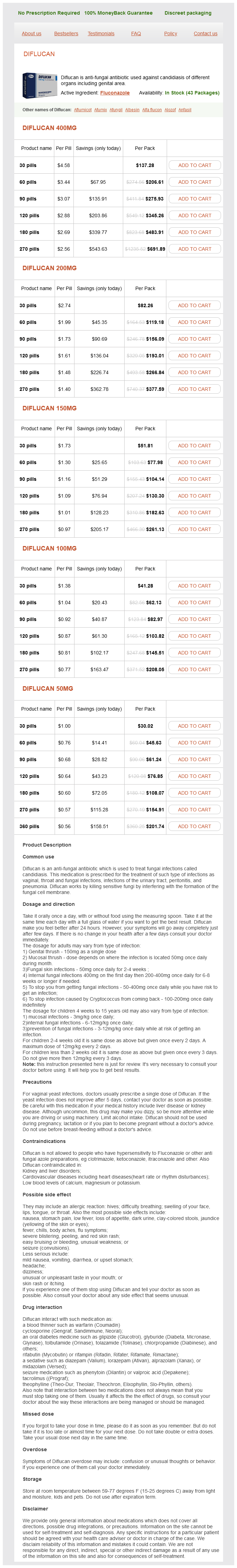
Fluconazole Dosage and Price
Diflucan 400mg
- 30 pills - $137.28
- 60 pills - $206.61
- 90 pills - $275.93
- 120 pills - $345.26
- 180 pills - $483.91
- 270 pills - $691.89
Diflucan 200mg
- 30 pills - $82.26
- 60 pills - $119.18
- 90 pills - $156.09
- 120 pills - $193.01
- 180 pills - $266.84
- 270 pills - $377.59
Diflucan 150mg
- 30 pills - $51.81
- 60 pills - $77.98
- 90 pills - $104.14
- 120 pills - $130.30
- 180 pills - $182.63
- 270 pills - $261.13
Diflucan 100mg
- 30 pills - $41.28
- 60 pills - $62.13
- 90 pills - $82.97
- 120 pills - $103.82
- 180 pills - $145.51
- 270 pills - $208.05
Diflucan 50mg
- 30 pills - $30.02
- 60 pills - $45.63
- 90 pills - $61.24
- 120 pills - $76.85
- 180 pills - $108.07
- 270 pills - $154.91
- 360 pills - $201.74
Occasionally it takes on a ring or annular distribution fungus wiki fluconazole 400 mg discount, extensively infiltrating the ciliary region. Malignant melanoma of the ciliary body is less common than that of the choroid; the treatment and prognosis are similar, but surgical excision is an option for smaller tumours. Epithelial hyperplasia of the ciliary processes is a small adenoma, that is not uncommon in old eyes. Rarely, Malignant Melanoma of the Choroid Pathology this arises from the outer layers of the choroid. The neurosensory retina remains in contact with the tumour at the summit, but is detached from the choroid at the sides, the intervening space being filled with exudative fluid. The growth may be in any location, and the fluid may sink down to the lowest part of the eye, forming a detachment isolated from that over the tumour, but with continuing growth the retina becomes more and more detached, until no part remains in situ. The tumour may fill the globe before perforating the sclera, or this may occur relatively early along the perivascular spaces of the vortex veins or ciliary vessels. The lymph nodes are not commonly affected, but metastases occur abundantly in the liver and elsewhere. The cells are usually spindle shaped; they may also be cylindrical or palisade-like, arranged in columns or around blood vessels, or even resemble endothelial cells in appearance; most tumours are mixed-celled. Spindle A-predominance of slender spindles with flattened nucleus and no nucleolus 2. Spindle B-predominance of larger spindles with round/ oval nucleus and prominent nucleolus 3. Clinical Features In adults, choroidal melanoma is the commonest intraocular malignant tumour. B-scan ultrasonography demonstrating a nodular extrascleral extension along the base of a relatively flat intraocular tumour (a, arrows). The extrascleral extension of the tumour should be differentiated from the extraocular muscles, which have flat configuration and appear to separate from the sclera when traced posteriorly corresponding to the normal anatomic location of the muscle (b, arrows). The growth is usually pigmented but is occasionally unpigmented, a distinction which is relatively unimportant. The pigment is chiefly melanin, but haematogenous pigmentation occurs after haemorrhages. Peripherally located tumours usually attain a considerable size, and cause a retinal detachment before the patient becomes symptomatic. It is of the utmost importance that the cause of a detachment of the retina should be identified in all cases. If a retinal detachment is accompanied by raised intraocular pressure, a growth may be diagnosed almost with certainty. A simple detachment shows numerous folds and undulations can be seen to travel over the surface when the eye moves. Patches of pigment upon the rounded part support the diagnosis of a tumour, but pigmentary disturbance, more particularly at the periphery, is not uncommon in a simple detachment. Commonly an orange pigment, lipofuscin, is deposited on the surface of the tumour. A simple detachment of the non-exudative type always has a hole or tear in the retina somewhere; if it can be found it is the most positive evidence that a growth is probably not present. Rarely a dual circulation develops and a system of blood vessels having an entirely different mode of distribution from the retinal vessels can be made out between the latter; this is the most positive evidence of a growth, but it is only occasionally seen. A very small, round detachment in the macular region or upper part of the globe is almost certain to be due to a tumour of the choroid. If the detachment is anterior, transillumination will afford assistance in diagnosis; a simple detachment is transparent, a choroidal growth opaque. The cause of the glaucoma in some cases is the forward movement of the lens and iris due to posterior pressure, so that the angle of the anterior chamber becomes blocked and a sudden rise in tension is precipitated. In other cases, particularly those of early onset, obstruction to the venous outflow from the eye is a possible explanation, the tumour being, in some instances, so situated as to press upon a vortex vein. In the differential diagnosis, two other tumours must be kept in mind, particularly in the early stages. A choroidal naevus appears as a bluish patch with somewhat feathered edges, usually about the size of the optic disc and situated near the posterior pole of the eye. It is congenital and symptomless but like naevi elsewhere, may occasionally assume malignant characteristics. A cavernous haemangioma of the choroid, another rare tumour of congenital origin and of exceedingly slow growth, is also usually situated near the disc. It has a greyish hue and indefinite margins and often causes an exudative retinal detachment. The differential diagnosis also includes posterior scleritis which may be difficult to distinguish from a malignant melanoma of the choroid if localized posteriorly. Diagnosis Investigations for the diagnosis of choroidal melanoma include B-scan ultrasonography, radio-isotope uptake studies, especially when the media are opaque, and fluorescein angiography. Ultrasonography permits the delineation of the overlying retinal detachment and provides details of any underlying tumour mass. Ultrasonographic measurements of the dimensions of the tumour, particularly the height or thickness and maximum horizontal diameter, are helpful in planning treatment. Radioactive tracers: Neoplastic tissue has an increased rate of phosphate uptake and retains the isotope longer than non-neoplastic tissue. The range of b-rays is small, about 23 mm on an average with a maximum of 78 mm, and this restriction makes the technique of the measurement of 32P uptake difficult. A solid-state detector is capable of distinguishing clearly between the majority of benign and malignant intraocular lesions.
© 2025 Adrive Pharma, All Rights Reserved..