General Information about Casodex
One of the benefits of utilizing Casodex within the treatment of prostate cancer is that it's taken orally, as a tablet. This makes it a convenient and non-intrusive treatment option for patients. It also has a comparatively low risk of side effects in comparison with other anti-androgen drugs.
However, like any treatment, Casodex could cause unwanted effects in some sufferers. The commonest unwanted side effects embrace scorching flashes, breast tenderness or enlargement, and decreased libido. In rare cases, it can also trigger liver issues, so common liver perform exams are beneficial while taking the medicine.
Androgens, similar to testosterone, are male hormones that promote the growth and function of the male reproductive system. In some cases, they will additionally stimulate the expansion of certain kinds of cancer cells, corresponding to those within the prostate gland. Casodex works by binding to those androgen receptors and stopping the androgens from attaching to them. It does not decrease the degrees of androgens in the body, however it does disrupt their exercise, resulting in a regression of the prostate tumor.
Casodex, also recognized as bicalutamide, is a nonsteroidal anti-androgen treatment used within the remedy of prostate most cancers. It belongs to a category of medicine referred to as racemic mixtures, which comprise equal amounts of two different types of the same compound.
In conclusion, Casodex is a nonsteroidal anti-androgen medication that effectively treats prostate most cancers by blocking androgen receptors and inhibiting the growth of most cancers cells. It does not affect hormone ranges within the body and can be used as a monotherapy or in combination with other therapies such as radiation therapy. With its convenient oral form and relatively low threat of unwanted effects, Casodex is a priceless possibility within the fight towards prostate most cancers.
The effectiveness of Casodex has been demonstrated in multiple medical trials. In a examine printed in the New England Journal of Medicine, it was discovered that Casodex as monotherapy considerably improved general survival charges in patients with advanced prostate cancer compared to a placebo. Another study confirmed that the addition of Casodex to radiation remedy improved survival rates in males with intermediate or high-risk prostate most cancers.
Casodex is primarily used as a medicine for monotherapy, meaning it's used as the main remedy for prostate most cancers. It can also be utilized in mixture with other treatments, corresponding to radiation therapy, to further goal the cancer cells. When used in mixture with radiation therapy, Casodex can improve the treatment�s effectiveness and lead to better outcomes for sufferers.
One of the principle advantages of utilizing Casodex is that it does not affect the endocrine system in any method. This implies that it does not affect the production of hormones, which might have undesirable unwanted side effects in some patients. Instead, the mechanism of action of Casodex lies in its ability to dam the androgen receptors within the body, notably these present in prostate cells.
Many prospective clinical trials have shown that either of the listed cephalosporins alone prostate cancer hip pain buy casodex online from canada, without doxycycline, will result in a clinical cure. For that reason, doxycycline administration could be reserved until the patient can take oral medication. Gynecologic Infection decttosurgical needle-tip excision, ayotherapy, or laser ablation. Another topical immune-modulating agent is a 15-percent sinecatechias ointment (Ve. Of other agents, trichloroacetic add and bichloroacetic add are proteolytic and are applied serially to warts. All topical treatments in this section can result in skin irritation, which at times may require treatment breaks. Thus, in general, treatment is selected based on clinical circumstances and patient and provider preferences (Werner, 2017). Importantly, no treatment option, even surgical excision, boasts 100-pcrcent clearance rates. Biopsy is coruidered for atypical or recalcitrant lesions, which may harbor concomitant intraepithelial neoplasia (Massad, 2011). External genital warts can devdop at sites in the lower reproductive tract, urethra, anus, or mouth. They arc usually asymptomatic but can be pruritic or painful depending on their size and location. Biopsy is not required unless coexisting neoplasia is suspected, the diagnosis is unclear, or the patient is imm. Condyloma acuminata may remain unchanged or spontaneously resolve, and the effi:ct of treaunent on future viral transmission is unclear. Recommended Treatment of External Genital Warts Patient-applied lmlqulmod 5% cream (Aldara). Apply a thin layer and rub until absorbed once daily at bedtime, three times a week, up to 16 wks. The area treated does not exceed 1ocm2, and the total volume of podofilox is limited to 05 mUd. If an excess amount is applied, the treated area is powdered with talc, baking soda, or liquid soap to remove unreacted acid. Of these, topical imiquimod is indfective and may cause severe application-site reactions (van der Woud. This mite is crab-shaped, and the female digs into the skin and remains there for approximately 30 days, elongat· ing her burrow. Sexual transmission is the most likdy cause of initial infection, but household contacts can become infected. A dda~ hypersensitivity reaction (type 4) to the mites, eye, and fcccs develops and results in crythematous papulcs, vesicles, or nodules in association with skin burrows. Definitive testing requires scraping across the burrow with a scalpd blade and mixing these fragments in immersion oil on a microscope slide. A thin layer is applied from the neck downward with special attention to pruritic areas and the hands, feet, and genital regions. Eight to 14 hours after application, a shower or bath is taken to remove the medication. Another option is a 200-µglkg sin· gle oral dose of ivermectin (Stromectol) given initially and then repeated 2 weeks later. Molluscum contagiosum is contagious until lesions resolve, and an incubation period of 2 to 7 weeks is typical. Alternativdy, material from a lesion can be collected on a swab, applied to a slide, and submitted for diagnostic staining. If these lesions become infected, antibiotics directed against skin flora may be necessary. Lice attach to the base of human hair with claws that vary in diameter between species. Pubic lice are usually sexually transmitted, whereas head and body lice may be traosmitted by sharing personal objects such as combs, brwhes, and clothing. Each female adult pubic louse lays approximately four eggs a day, which are glued to the base of hairs. Their attached eggs, termed nits, can be seen attached to the hair shaft away from the skin line as hair growth progres. Moreover, suspicious flecks on pubic hair or in clothing can be examined microscopically to see the characteristic louse. Lice depend on frequent human blood meals, and pubic lice must travel for new attachment sites. A single application is usually dfective, but a second dose is recommended within 7 to 10 days to kill new hatches. Also, ivermeotin, 250 µg/kg orally once, can be taken and then repeated in 2 weeks. These areas are best treated by applying petrolatum (Vaseline) with a cotton swab at night and washing it off in the morning. Bacteria ascend from the short, colonized urethra and easily enter the bladder and perhaps then the kidneys. Also, contributing to contamination, the warm moist vulva and rectum are both in close proximity.
In these cases prostate cancer va disability compensation casodex 50mg buy with amex, samples from voided urine, the vagina, or the cndoccrvix arc appropriate (Lillis, 2011). Many of these patients have antibodies to C trachomatis and/or N gono"hoeae (Sellors, 1988; Tjiam, 1985). At laparoscopy or laparotomy, affected women may have evidence of prior tubal infection such as adhesions or hydrosalpingcs, but for the most part, the fallopian tubes are grossly normal. Internally, however, tubes show flattened mucosal folds, extensive deciliation of the epithelium, and secrctory epithelial cell degeneration (Patton, 1989). For reproductive-aged women, pregnancy complications can be identified by serum or urine beta-human chorionic gonadotropin testing. In general, the delay between collection and reporting precludes its clinical utility. One or more of the following enhances diagnostic specificity; (1) oral temperature >38. Unfortunately, no single symptom is associated with a physical finding that is specific for this diagnosis. Accordingly, other possible sources of acute pelvic pain are considered and listed in Table 12-1 (p. Of findings, mucopurulent endocervicitis is common and is diagnosed visually and microscopically. If a woman has pelvic peritonitis secondary to bacteria and pus that has exuded from the fimbriated end of the fallopian tube into the pelvis, this rapid peritoneal movement usually causes a marked pain response. Tapping the posterior culde-sac with an examining finger will give the examiner similar information. In women with marked abdominal pain and tenderness, appreciation of upper reproductive tract organs during bimanual examination may be limited, and sonography is a primary imaging tool. However, with acute tubal inflammation, the tube swells, its lumen occludes distally, it distends, and its walls and endosalpingeal folds thicken. If color or power Doppler is applied, marked vascularity with low-impedance blood flow, which reflects hyperemia, is seen within thickened fallopian tube walls and septa (Molander, 2001; Romosan, 2013). If sonography does not lead to a clear diagnosis, computed tomography scanning may be useful and especially with right upper quadrant pain suggestive of perihepatitis (Kim, 2009). Transvaginal sonogram shows a pyosalpinx and both ovaries as part of a tuboovarian complex. Computed tomography Image shows a tortuous pyosalplnx arrow) adjacent to the uterus (Ut). Microorganisms frequently cultured from abdominal specimem include Escherkhia C<Jli and Blll:tmJiJes, Peptosmpt«ocr:t1S, and Strtp1«oa:w species (Landers, 1983). Tuboovarlan Abscess With infection, the inBamed and suppw:ative fallopian tube can adhere to the ovary. With abscess progression, further structural weakening may lead to abscess rupture and potentially life-threatening peritonitis. In addition to those qualities just described, the mass often shows surrounding i. Gynecologic Infection 71 Parenteral antimicrobial therapy is continued until the patient has been afebrile for at least 24 hours, preferably 48 to 72 hows. For th0&e not improved within 2 to 3 days of tteaunent, antimicrobial regimen modifu::ation is reaaonable. The plgtall catheter coils to remain In place to allow conotic therapy can be considered tinuous drainage. Radiologic drainage is minimally cent; two episodes, 20 percent; and three or more episodes, invasive and potentially avoids the higher risks aasociated with 40 percent Westrom, 1992). However, some criteria predict better n«dle aspiration or with pigtail catheter plac:ement and shortoutcome for certain patients (Table 3-10). During the 6rst 3 weeks after insertion, coagulopathy (American College of Radiology, 2018). Although gery is required, and goals are abscess drainage, necrotic tissue a provider may choose to remove the device, evidence supports acision, and peritoneal cavity irrigation. But, if a patient f:Ws to improve within 48 to toneal and other tissue planes to remove tissues-especially the 72 hours, the device is removed. However, a cul-do-sac, inu:rloop, or multiloculau:d absa:ss High fever is more likely to require drainage. Generalized peritonitis Failed outpatient therapy · Treatment of Acute Pelvic Noncompliant with medications Inflammatory Disease White blood cell count > 15,000/mm3 Nausea/vomiting precluding oral therapy the most successful patient outcomes follow early diagnosis and prompt, appropriate therapy. For women with outpatient treatment, one study showed that women treated as outpatients took 70 percent of prescribed doses, and for less than 50 percent of their outpatient treatment days (Dunbar-Jacob, 2004). Thus, if patients are treated as outpatients, an initial parenteral dose may be beneficial. Women treated as outpatients are reevaluated in approximately 72 hours by phone or in person. Alternatively, if the N gentamycin/clindamycin regimen is used, transition to a 14-day oral agent may involve clindamycin orally four times daily or doxycycline 100 mg twice daily. Pelvic infection and abscess are rare, even in those identified to harbor the bacteria. With infection, findings include fever, weight loss, abdominal pain, and abnormal vaginal bleeding or discharge. In the absence of symptoms, the incidental finding of Actinomyces in a Pap sample may be managed conservativdy. If signs or symptoms of infection do develop in a woman who harbors Actinomyces, the device is removed and antimicrobial therapy is instituted. Although rare in the United States, salpingitis and endometritis can originate from pulmonary tuberculosis. Mycobacterium tubm:uwsis is thought to be blood-borne, but ascension may still be a possible route.
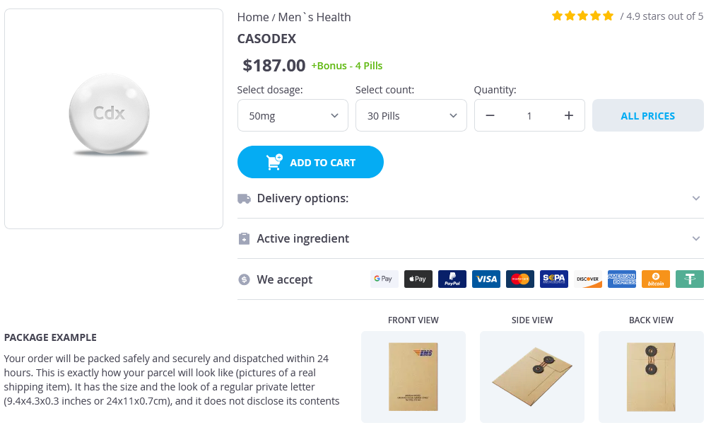
Casodex Dosage and Price
Casodex 50mg
- 30 pills - $207.78
Other ablative techniques such as microwave ablation prostate oncology specialists nj cheap 50 mg casodex with visa, laser ablation and high-intensity ultrasound ablation have been used but are considered experimental. There are multiple bilateral renal cysts with a malignant looking lesion in the upper pole of the left kidney and possibly the lateral aspect of the lower pole of the right kidney. Given this and the history of cerebellar surgery I suspect that he has von HippelLindau disease. This is an autosomal dominant disorder with an incidence of 1:36,000 live births which affects males and females equally. The average age of the patient at diagnosis is 26 years and is characterised by the development of various benign and malignant tumours and cysts. The major tumours and cysts are haemangioblastoma in the central nervous system, retinal haemangioblastoma, phaeochromocytoma, renal cell carcinoma, renal cysts, pancreatic neuro-endocrine tumours, and pancreatic and epididymal cystadenomas. However, diligent surveillance enabling early treatment interventions can increase life expectancy. Pazopanib is an oral angiogenesis inhibitor and is comparable to Sunitinib in outcomes, but with better quality of life. Although, not a uniform policy, one approach to medical therapy is initially to stratify patients according to favourable, intermediate, and poor risk, depending upon whether they possess zero, one or two or more of the following risk factors: Karnofsky performance status 80%, anaemia, elevated serum calcium, absence of prior nephrectomy, and elevated lactate dehydrogenase [13]. Poor risk patients, especially those with non-clear-cell histology, should initially be considered for treatment with Temsirolimus. Importantly, with several ongoing clinical trials in progress, treatment options are likely to change. Kidneys usually develop a spectrum of small benign cysts through to large renal cell cancers. Smaller lesions are difficult to evaluate but a cut-off at 23 cm is thought to represent increasing risk of malignant transformation. Cryo-ablation or radio-frequency ablation should be considered for treating smaller lesions (<3 cm). Ultimately as the disease progresses the patient may need renal replacement therapy. The evidence for the role of transplantation and subsequent immuno-suppression in this population of patients is limited. There are a number of other cancer susceptibility syndromes that are associated with an increased risk of renal cancer. The mean age of diagnosis is 40 years and metastatic renal cancer can present in the teens. The most common type of tumour is an unusual hybrid oncocytic tumour (mixed oncocytoma and chromophobe). Tuberous sclerosis complex is an autosomal dominant genetic disorder characterised by the formation of hamartomas in multiple organs including the brain, kidney, skin and lung. This leads to neurological disorders including epilepsy, mental retardation and autism as well as dermatological manifestations such as facial angiofibromas. The relevant radiological features include calcification, septations, irregular margins, solid elements and evidence of contrast enhancement. Uniformly high-attenuation lesions <3 cm in size, with sharp margins without enhancement. The cyst may contain calcification, which may be nodular and thick, with no contrast enhancement. This category also includes totally intrarenal, nonenhancing, high attenuation renal lesions >3 cm. These are indeterminate cystic masses with thickened irregular walls Surgery or active surveillance as or septa with enhancement. Enhancement suggests the presence of vascular tissue or communication with the collecting system. It is measured by the difference in Hounsfield units pre- and post-contrast and can point towards a malignant diagnosis (an approximate increase in enhancement of 15 or more Hounsfield units is considered significant). What is the difference between autosomal dominant polycystic kidney disease, autosomal recessive polycystic kidney disease and acquired renal cystic disease Diagnosis is often made in utero with the development of bilateral enlargement of renal parenchyma which is replaced by radially orientated cysts. Oligohydramnios may occur and, if severe, termination is often considered in the second trimester. Management is supportive including good blood pressure control, dialysis and consideration of transplantation. Uraemic toxins are implicated and cyst regression after transplantation and recurrence after transplant failure are seen. A spectrum of renal adenoma to carcinoma is seen with a 3 cm cut-off usually considered for a malignant diagnosis. Unilateral multicystic dysplastic kidney has an incidence of 1:25001:4000 and presents as an incidental finding or as an irregular flank mass. Where inherited, it is an autosomal dominant disorder, although sporadic cases are more common. There is proximal ureteric atresia secondary to ureteric bud and metanephric mesenchymal defects. Most unilateral cases are undetected at birth, and involution commonly occurs in early childhood which may account for the recognised association with renal agenesis. Multilocular cysts tend to be bulky with thick capsules containing highly echogenic septae with loculi sometimes containing debris suggesting solid elements. Distribution is bimodal, with 2:1 male:female predominance under 4 years, and 8:1 female predominance above 30 years. Children present with an asymptomatic flank mass, adults with abdominal pain or haematuria. A 43-year-old female presented acutely to the accident and emergency with loin pain.
© 2025 Adrive Pharma, All Rights Reserved..