General Information about Betapace
Betapace belongs to a category of medicines often identified as antiarrhythmics, which work by controlling the electrical impulses that trigger the center to beat irregularly. It does this by blocking particular channels in the coronary heart which are liable for transmitting these impulses. By doing so, it helps in restoring a traditional coronary heart rhythm, thereby lowering the chance of problems.
One of the significant benefits of Betapace is that it is obtainable in both immediate-release and extended-release formulations, making it handy for patients with completely different wants. The immediate-release formulation is used for rapid remedy and is often taken two or three times a day, while the extended-release choice is taken only as soon as a day, making it extra appropriate for long-term use.
In some instances, Betapace is in all probability not appropriate for people with pre-existing heart circumstances, liver or kidney illness, or a recognized allergy to sotalol. Pregnant or breastfeeding ladies must also use Betapace with caution and only under medical supervision.
As with any medicine, Betapace additionally comes with some potential side effects that sufferers should pay attention to. These include dizziness, headache, fatigue, nausea, and diarrhea. While most of these unwanted effects are mild and don't require medical attention, in some uncommon instances, extra extreme side effects, corresponding to chest ache, issue respiration, or swelling of the face, can occur. If any of these signs are experienced, it is crucial to seek medical help immediately.
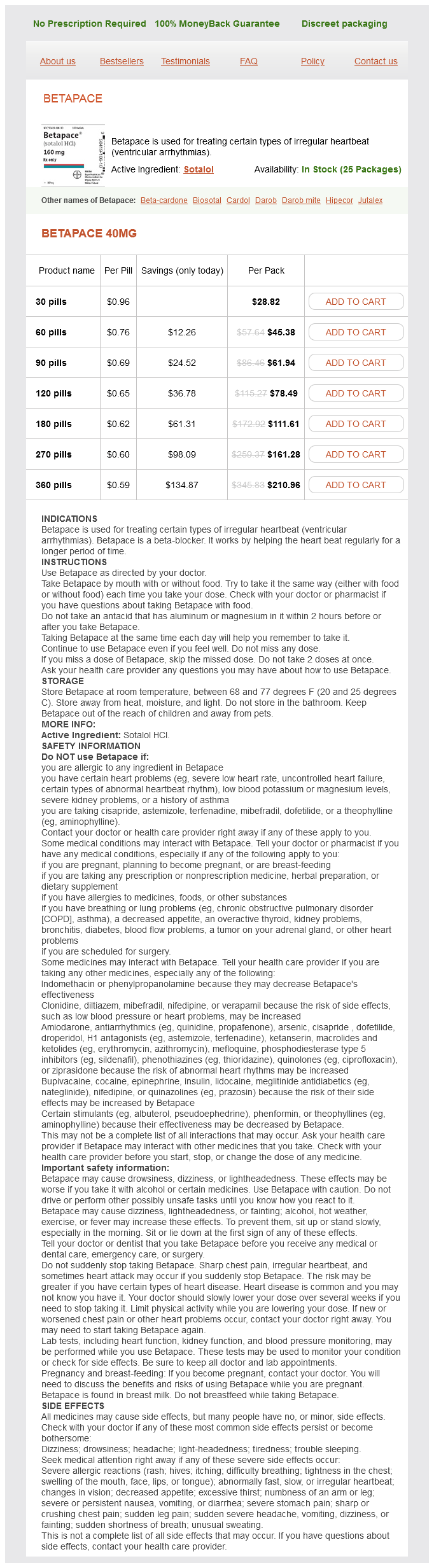
Betapace, also recognized by its generic name, sotalol, is a medicine used within the therapy of certain kinds of irregular heartbeats, medically often recognized as ventricular arrhythmias. These conditions can happen as a result of numerous causes, such as heart disease, certain drugs, or electrolyte imbalances. If left untreated, ventricular arrhythmias can be doubtlessly life-threatening, making Betapace a significant medicine for people who endure from these situations.
In conclusion, Betapace is a vital medicine for those affected by irregular heartbeats, notably sustained ventricular tachycardia and ventricular fibrillation. Its effectiveness in controlling these situations and its handy dosing choices make it a preferred selection for so much of doctors and patients. However, like all treatment, it's essential to make use of Betapace as directed and to monitor for any potential unwanted facet effects. With proper utilization and monitoring, Betapace can help individuals with ventricular arrhythmias live a healthier and more snug life.
The dosage of Betapace may range depending on the severity of the condition, the affected person's age, and different underlying health situations. It is crucial to observe the prescribed dosage and not to regulate it with out consulting a doctor first. A sudden change in dosage can lead to serious unwanted effects, including a sudden fast heartbeat, dizziness, or fainting.
Betapace is normally prescribed for a selected sort of ventricular arrhythmia referred to as sustained ventricular tachycardia, where the center beats at an abnormally quick tempo for an extended interval. It can be used to treat a extra extreme type of arrhythmia often identified as ventricular fibrillation, the place the center beats with chaotic, uncoordinated electrical impulses, causing it to quiver instead of pumping blood effectively.
Betapace also can interact with different drugs, together with blood strain medications, sure antibiotics, and antidepressants. It is crucial to tell the physician about all of the drugs and dietary supplements one is at present taking earlier than starting Betapace to avoid potential interactions.
When necessary blood pressure systolic purchase discount betapace on line, surgical excision and/or radiofrequency catheter ablation therapy was effective (103). The chest radiograph, however, often shows cardiomegaly and pulmonary edema in the critically ill neonate and infant (7,19,40,74,76,77,78,79). The 2-D and 3-D echocardiographic characteristics of cardiac rhabdomyomas are highly echogenic, multiple, well-circumscribed intramural or intracavitary nodules occurring anywhere within the heart. Rhabdomyomas also may be visualized as either single pedunculated intracavitary or intramural masses (23,40,41,42,78,85,97,103). Moreover, rhabdomyomas do not have interspersed, distinct, echogenic regions consistent with calcification or fibrosis. Multiple tumors are highly indicative of rhabdomyomas both in the fetus and neonate (15,16,80,81). Cardiac rhabdomyomas are closely associated with the syndrome of tuberous sclerosis. Tuberous sclerosis is a complex disorder that is transmitted as an autosomal dominant trait of variable expression (104,105,106,107,108,109,110,111,112,113). The incidence of tuberous sclerosis has been reported to be 1 in 6,000; two-thirds of cases are sporadic resulting from mutation (81). In an autopsy series, 31% of patients with tuberous sclerosis were reported to have had rhabdomyomas (8). Fifty percent of patients with tuberous sclerosis had echocardiographic evidence of tumor involvement (15,85). Fetal studies indicate that 52% to 79% of all fetuses with cardiac tumors have tuberous sclerosis (15,16,80). The incidence of tuberous sclerosis is as high as 59% to 80% in patients with confirmed fetal rhabdomyomas. Although not invariably, multiple rhabdomyomas are more consistent with the diagnosis of tuberous sclerosis than a singular tumor, with one report indicating that in the fetus or newborn with multiple ventricular tumors, 95% had tuberous sclerosis (16). However, in the latter report, 23% of fetuses or neonates with a singular tumor had tuberous sclerosis as well. In addition to cardiac involvement, tuberous sclerosis can affect almost all organ systems, including the brain, kidney, pancreas, retina, and skin (104,105,107,108,109). Even in severely affected patients, clinical manifestations such as Shagreen patches, adenoma sebaceum, seizures, and mental retardation may not become evident until later in life (105,107,108,109,110,111,112). Recently cardiac fat containing lesions have been reported in adults with tuberous sclerosis which are distinct from rhabdomyomas and are seen in one third of adolescent/ adult patients. There appears to be a higher incidence of abdominal angiomyolipomas in such patients. Neonates with tuberous sclerosis, however, may have no manifestations of this syndrome other than cardiac tumors (79,90,94,95,108). Furthermore, a negative family history does not preclude the diagnosis since 50% of cases of tuberous sclerosis may be spontaneous mutations. Recent advances in genetic testing may elucidate more fully the inheritance of tuberous sclerosis. These loci code for proteins, hamartin and tuberin, that have a tumor-suppressor function (113). Prompt surgical excision is indicated for life-threatening hemodynamic compromise or arrhythmias (38,39,76,79) and is required in 23% of patient series (97). Partial excision may provide significant relief of inflow or outflow tract obstruction when attempts at complete resection would severely damage the remaining myocardium P. The Ross procedure has been proposed when the aortic valve is severely compromised (115). Surgical intervention may be highly successful, without compromising either myocardial or valvular function, in patients with single intracavitary rhabdomyomas (40,84,100). Others have reported successful radiofrequency ablation therapy in selected patients with rhabdomyomas and supraventricular tachycardia (116). However, even very large rhabdomyomas may significantly regress in size or disappear completely without intervention (22,38,39,90,100). Unoperated patients may have minimal cardiac signs or symptoms several years later (90). Therefore, the presence of rhabdomyomas alone, without life-threatening hemodynamic instability or arrhythmias, should not be an absolute indication for surgery (18,19,38,39,90,100). Fibromas Fibromas are generally reported as the second most common primary cardiac tumor in the pediatric age group (Videos 72. Although fibromas have recently been reported in utero (14,15) and in patients younger than 1 month of age (34,35,36,37,121,122,123,124), they are found much less commonly than rhabdomyomas in this age group. Sudden death has been attributed to cardiac fibromas in pediatric patients of all ages (31,124,125). These tumors have been associated with Gorlin syndrome, which includes multiple nevoid basal cell carcinomas, cysts of the jaw, and diffuse skeletal abnormalities (126). Cardiac fibromas also have a rare association with familial adenomatous polyposis and its subtype Gardner syndrome (127,128). Cardiac fibromas are predominantly single, white, firm, nonencapsulated, intramural tumors that involve the left ventricular free wall or interventricular septum (32,37,38,39,40,41,42,43,44,45,46,47,48,49,50,51,52,53,54,55,56,57,58,59,60,61,62,63,64,65,66,67,68,69,70,71,72,73,74,75,76,77,78,79,80,81,82,83,84,85,86,87,88,89,90,91,92,93 these tumors often are located at the left ventricular apex. Less frequently, fibromas can be multiple and invade the right ventricular free wall, atrial septum, or atrial free wall (32,35,36,37,123,124). Extensive intramural fibromas can encroach and obliterate the intracavitary space (35,36,37,123,124,129).
Safety of cardiac catheterization at a center specializing in the care of patients with pulmonary arterial hypertension arrhythmias 40 mg betapace buy otc. Impact of pulmonary vascular stiffness and vasodilator treatment in pediatric pulmonary hypertension: 21 patient-specific fluid-structure interaction studies. Pulmonary vascular stiffness: measurement, modeling, and implications in normal and hypertensive pulmonary circulations. Computational Simulation of the pulmonary arteries and its role in the study of pediatric pulmonary hypertension. Extraction of pulmonary vascular compliance, pulmonary vascular resistance, and right ventricular work from single-pressure and Doppler flow measurements in children with pulmonary hypertension: a new method for evaluating reactivity: in vitro and clinical studies. Noninvasive Doppler tissue measurement of pulmonary artery compliance in children with pulmonary hypertension. The prognostic value of pulmonary vascular capacitance determined by Doppler echocardiography in patients with pulmonary arterial hypertension. Relationship of pulmonary arterial capacitance and mortality in idiopathic pulmonary arterial hypertension. Pulsatile haemodynamic parameters are predictors of survival in paediatric pulmonary arterial hypertension. Noninvasive methods for determining pulmonary vascular function in children with pulmonary arterial hypertension: application of a mechanical oscillator model. Evidence for the association of unexplained pulmonary hypertension in children with the major histocompatibility complex. Pediatric pulmonary arterial hypertension and hyperthyroidism: a potentially fatal combination. Bone morphogenetic protein receptor 2 mutations in adults and children with idiopathic pulmonary arterial hypertension: association with thyroid disease. Autoimmune thyroid disease in children and adolescents with idiopathic pulmonary arterial hypertension. Plasma brain natriuretic peptide as a prognostic indicator in patients with primary pulmonary hypertension. Prognostic value of B-type natriuretic peptide in children with pulmonary hypertension. Prognostic factors in pediatric pulmonary arterial hypertension: a systematic review and meta-analysis. Plasma proteomics of differential outcome to long-term therapy in children with idiopathic pulmonary arterial hypertension. Circulating fibrocytes are increased in children and young adults with pulmonary hypertension. Circulating myeloid-derived suppressor cells are increased and activated in pulmonary hypertension. Evaluation of circulating proteins and hemodynamics towards predicting mortality in children with pulmonary arterial hypertension. Pathophysiological and metabolic manifestations of pulmonary vascular disease in children. Reversal of pulmonary hypertension associated with plexiform lesions in congenital heart disease: a case report. Interstitial lung disease associated with surfactant protein B and C deficiencies. Surfactant protein C mutations are the basis of a significant portion of adult familial pulmonary fibrosis in a dutch cohort. Intravascular ultrasonic characteristics and vasoreactivity of the pulmonary vasculature in children with pulmonary hypertension. Magnification pulmonary wedge angiography in the evaluation of children with congenital heart disease and pulmonary hypertension. Pharmacologically induced pulmonary vasodilatation in children and young adults with primary pulmonary hypertension. Lower airway obstruction, bronchial hyperresponsiveness, and primary pulmonary hypertension in children. Primary pulmonary hypertension in children may have a different genetic background than in adults. Somatic chromosome abnormalities in the lungs of patients with pulmonary arterial hypertension. Serotonin transporter polymorphisms in familial and idiopathic pulmonary arterial hypertension. Isolated atrial septal defect with pulmonary vascular obstructive diseaselong-term follow-up and prediction of outcome after surgical correction. Preoperative pulmonary hemodynamics and assessment of operability: is there a pulmonary vascular resistance that precludes cardiac operation Comparison of hyperventilation and inhaled nitric oxide for pulmonary hypertension after repair of congenital heart disease. Pulmonary arterial hypertension and congenital heart disease: targeted therapies and operability. Measured versus estimated oxygen consumption in ventilated patients with congenital heart disease: the validity of predictive equations. Validity of the LaFarge equation for estimation of oxygen consumption in ventilated children with congenital heart disease younger than 3 yearsa revisit. Inhaled nitric oxide to test the vasodilator capacity of the pulmonary vascular bed in children with long-standing pulmonary hypertension and congenital heart disease. Combined effects of nitric oxide and oxygen during acute pulmonary vasodilator testing.
Betapace Dosage and Price
Betapace 40mg
- 30 pills - $28.82
- 60 pills - $45.38
- 90 pills - $61.94
- 120 pills - $78.49
- 180 pills - $111.61
- 270 pills - $161.28
- 360 pills - $210.96
The Lungs Pulmonary situs is determined by the sidedness of the morphologic right and left lungs blood pressure stroke range discount betapace online american express. In contrast, a morphologic left lung is characterized by a pulmonary artery that courses over the main bronchus and posterior to the upper lobe bronchus. In the setting of pulmonary isomerism (both lungs having the same morphology), however, the ratio approaches unity, because the lengths of the two main bronchi are similar. Pathologic specimen (A) and drawing (B) illustrate normal tracheobronchial branching pattern as observed anteriorly. This anatomic feature predicts pulmonary visceral situs based on measurements of the bronchial length. For example, atria and ventricles should be described both by their morphology and their right or left sidedness. Mesocardia is characterized by a vertical midline septum with side-by-side ventricles. Rarely, the ventricular septum is horizontal rather than vertical and results in superoinferior (over-and-under or "upstairs downstairs") ventricles. After the morphology and position of the cardiac segments are determined, the manner in which they connect to one another is evaluated. The difficulties encountered in the accurate definition of atrial situs based on atrial anatomy alone emphasize the importance of accurate determination of systemic and pulmonary venous connections, as described in Tables 51. This determination is particularly important for surgical management and should be a routine part of any imaging study. Although these basic combinations do not include all possible variations in ventricular and great artery spatial relationships, the important aspects of atrioventricular and ventriclegreat artery connections are demonstrated. Ventricular Positions and Morphology Complete assessment of this segment includes an assessment of the cardiac baseapex axis to describe the three basic possibilities of levocardia, dextrocardia, or mesocardia as detailed previously. As covered elsewhere in this chapter, an evaluation of this segment should incorporate descriptions of the ventricular locations, relationships, function, and morphology, as well as the anatomy of the ventricular septum and any septal defects. Great Artery Relations the great arterial connections, the spatial relationships between the arteries and their subsequent distribution/branching patterns, must be determined as described in Table 51. Eight basic types of great artery relationship are possible based on the aortic and pulmonary valve positions at the level of the semilunar valves. Directly posterior aorta this type of segmental approach to complex cardiovascular malformations allows prediction of all possible cardiac conditions that could be encountered. Its flexibility depends upon the sequential addition of each cardiac segment and relevant connections, accounting for all possible combinations of atrial, ventricular, and great arterial anomalies. Cardiac Imaging: Use of the Segmental Approach Transthoracic echocardiography is a convenient and noninvasive technique that can provide most of the clinically relevant information concerning the cardiovascular anatomy and physiology, especially in young patients. We will primarily use echocardiographic examples to illustrate the segmental approach, but the concepts are applicable to , and should be used in all tomographic imaging modalities. The imaging approach to patients with cardiac malpositions should follow the same sequential segmental pattern used by the anatomist/pathologist as outlined earlier in this chapter and in many reports (20,21,22,23). The following section and accompanying images of normal and malpositioned hearts are included to illustrate the tomographic and three-dimensional anatomy associated with these malformations and to assist the reader in correlating the imaging findings with anatomic examples of these complex hearts. As previously described, situs ambiguus has been used to describe a wide spectrum of anomalies, including the visceral P. The visceral situs is considered "ambiguous," because it does not conform to the classical patterns of situs solitus or inversus. Nearly all possible combinations of abdominal organ and great vessel location have been reported (19,22). The term visceral situs ambiguus does not mean that the positions of the abdominal organs cannot be determined. Subcostal short-axis scan of the abdomen provides right-to-left spatial orientation. The obvious echocardiographic finding in patients with asplenia is the inability to demonstrate a spleen. The spleen, when present, is always located posterolateral to the stomach (7,11,19,24). Routine transthoracic echocardiography can easily determine splenic status by locating the stomach and interrogating the area posterior and lateral to the stomach. The splenic tissue may have a denser echocardiographic appearance than the liver and its comma-shaped curvilinear splenic vein may be identifiable. When this echocardiographic finding is associated with the presence of HowellJolly bodies on the peripheral blood smear, the diagnosis of asplenia can be made with 100% confidence. The liver will frequently have two lobes that are relatively equal in size and resemble morphologic right lobes. A: Asplenia: subcostal short-axis horizontal-plane image of the upper abdomen in a patient with situs ambiguus and asplenia. B: Polysplenia: subcostal short-axis horizontal-plane image of the abdomen in a patient with situs ambiguus and polysplenia. C: Long-axis image in the same patient demonstrating the aorta and the azygos vein. Patients with polysplenia will have multiple, separate spleens positioned posterior to the stomach. As with asplenia, multiple or multilobulated spleens can be readily detected by P.
© 2025 Adrive Pharma, All Rights Reserved..