General Information about Amitriptyline
It is primarily used to deal with depression, nevertheless it may additionally be used for different situations similar to chronic ache, nervousness, and insomnia.
In addition to treating depression, amitriptyline has also been found to be useful in managing continual ache. This is because it blocks the reuptake of serotonin and norepinephrine, two neurotransmitters which are concerned in ache perception. By increasing the levels of those chemical compounds in the mind, amitriptyline can provide aid from persistent pain conditions similar to fibromyalgia, neuropathic pain, and migraines.
As with any treatment, there are potential unwanted side effects that will happen when taking amitriptyline. Common unwanted facet effects include dry mouth, constipation, blurred imaginative and prescient, dizziness, and drowsiness. It is important to debate any unwanted effects with a health care provider as they could be able to regulate the dosage or switch to a special medication.
Another frequent use for amitriptyline is within the treatment of tension problems. Due to its ability to increase serotonin ranges, it can help reduce signs of tension such as racing ideas, restlessness, and panic attacks. It is commonly prescribed for generalized anxiety dysfunction, social nervousness disorder, and post-traumatic stress dysfunction.
When taking amitriptyline, it is important to observe the prescribed dosage and to not cease taking the medication abruptly. Suddenly stopping the treatment can result in withdrawal signs such as nausea, headache, and irritability. Additionally, it could take a number of weeks before the total results of amitriptyline are felt, so you will need to be affected person and continue taking the medication as directed.
Amitriptyline may also be used off-label to treat different circumstances such as insomnia. Its sedative results might help people fall asleep and stay asleep, making it useful for these struggling with sleep issues. It is commonly prescribed at lower doses for this objective compared to its use in treating despair or nervousness.
In conclusion, amitriptyline is a widely used treatment for the remedy of despair, continual pain, nervousness, and insomnia. It works by rising the degrees of certain chemical compounds in the mind that play a role in regulating mood and ache perception. While there may be potential unwanted side effects, this treatment has been confirmed to be effective in improving the quality of life for individuals battling psychological health issues. As always, it is necessary to seek the advice of with a doctor and comply with their guidance when considering any treatment.
One of the primary uses of amitriptyline is for the remedy of depression. It is often prescribed as a first-line treatment for individuals with moderate to extreme despair. This is as a outcome of it has been found to be extremely effective in relieving signs corresponding to feelings of hopelessness, lack of interest in beforehand enjoyable actions, and adjustments in urge for food and sleep patterns.
Amitriptyline just isn't really helpful for everyone and should not be taken without the supervision of a physician. It may work together with different medicines or situations, so it is necessary to disclose all medications and medical historical past to a healthcare supplier earlier than beginning remedy.
Amitriptyline has been around since the Sixties and continues to be commonly prescribed today due to its effectiveness in treating a wide range of psychological well being issues. This medication works by altering the balance of sure chemical compounds in the brain which are responsible for regulating temper and feelings.
Treatment was well tolerated; toxicities were quickly solved without dose reductions or interruptions anxiety and chest pain cheap amitriptyline online mastercard. Pediatric Nephrology, Hypertension and Pheresis, Washington University School of Medicine & St. There remains an absence of a standard frontline treatment approach in the real world setting. Survival endpoints estimated by Kaplan-Meier method and compared by log rank test. A minority had graft involvement or rejection (19% each) at the time of diagnosis. These data confirm the findings in the preclinical studies, suggesting these unique, non - chemotherapy based combinations exhibit a selectivity in one lineage of lymphoma over another. We will share the evolving biological rational and early preclinical and clinical experience which warrants further study of these potentially paradigm changing approaches. Concomitant chemoradiotherapy and sequential chemotherapy and radiotherapy have similar response rates and survivals. Patients were randomly assigned to receive either P-Gemox+thalidomide regimen (Group A: Pegaspargase 2000U/m2; im d1, Gemcitabine 1000mg/m2; iv drip, d1, d8. Overall response during induction in Group A and B was similar in both groups, were 64. Group B was better tolerated than Group A, with lower rates of agranulocytosis, thrombocytopenia and infections. Two patients died of severe acute renal failure and sepsis at the first cycle, and one patient died of sepsis at the third cycle. For advanced or relapsed patients, both regimen showed unsatisfied survival outcome. There is established or presumed evidence that the resulting functional deregulations represent pathogenic mechanisms contributing to induce or maintain some attributes of the malignant phenotype. The immunotherapy of cancer has also made significant progress in the past decade. Improved understanding of cancer immunology increases insights into the mechanism of tumor response to immune cells, elucidating the therapeutic role of immunity in cancer and fueling an expanding array of new therapeutic agents. Together, immune dysregulation plays an important role on disease progression and may become potential therapeutic targets. Due to its rarity and the heterogeneity of subtypes, large, prospective, and randomized trials comparing different treatment approaches are still lacking. In recent years, early consolidation with high dose chemotherapy and stem cell rescue has been adopted in many Institutions, with promising results. In any case, the risk of relapse remains quite high and relapsed or refractory patients have been shown to have a very dismal outcome. For those patients in need of salvage therapy, current data confirm the unmet need for better treatment. However, a trend towards greater response and survival in those treated with single agents, while maintaining the ability to bridge to transplantation, is emerging. At present, 60 Institutions from 18 different countries already joined the project. Disclosures: Federico, M: Research Funding: Roche, Abbvie, Gador, Roemmers/Iclos, Libra, Innate, Nolver, Pfizer, Scienza Novartis, Thomas Jefferson University, Seattle Genetics Innate Pharma. Disclosures: Kwak, L: Consultant Advisory Role: Pepromene Bio, InnoLifes, Enzychem, Celltrion; Stock Ownership: Pepromene Bio, InnoLifes, Xeme BioPharma; Research Funding: Pepromene Bio. Sadelain Department of Medicine, Memorial Sloan Kettering Cancer Center, New York, United States; 2Michael G. Median age of the pts was 70 (range, 53-81), and median number of prior treatments was 5 (range, 2-17). Disclosures: Ramos, C: Consultant Advisory Role: Novartis, Celgene; Research Funding: Tessa Therapeutics. All patients received preceding lymphodepleting chemotherapy (cyclophosphamide 500 mg/m2 and fludarabine 30 mg/m2 daily for 3 days). The median number of prior therapies was 5 (range: 1-10), and 17 pts (89%) had high risk cytogenetics (17p deletion and/or complex karyotype and/or 11q abnormalities). Disclosures: Lymp, J: Employment Leadership Position: Juno Therapeutics, A Celgene Company; Stock Ownership: Juno Therapeutics, A Celgene Company. Li, D: Employment Leadership Position: Juno Therapeutics, A Celgene Company; Stock Ownership: Juno Therapeutics, A Celgene Company. Maloney, D: Honoraria: Janssen Scientific Affairs, Seattle Genetics, Roche/Genentech; Research Funding: Juno Therapeutics, GlaxoSmithKline. Turtle, C: Consultant Advisory Role: Juno Therapeutics, Nektar Therapeutics, Eureka Therapeutics, Precision Biosciences, Caribou Biosciences. Maloney, D: Honoraria: Janssen Scientific Affairs, Seattle Genetics, and Roche/Genentech; Research Funding: GlaxoSmithKline, and Juno Therapeutics, a Celgene/Bristol-Myers Squibb company. Turtle, C: Consultant Advisory Role: Caribou Biosciences, Eureka Therapeutics, Precision Biosciences, Aptevo, Humanigen, Juno Therapeutics, a Celgene/Bristol-Myers Squibb company, Kite, a Gilead Company, Nektar Therapeutics, and Novartis; Stock Ownership: Caribou Biosciences, Eureka Therapeutics, and Precision Biosciences; Research Funding: Juno Therapeutics, a Celgene/Bristol-Myers Squibb company, and Nektar Therapeutics. Two pts had grade 3 related events (neutropenic fever and cytopenia) after durvalumab treatment. Conclusions: Based on preliminary results, the combination of liso-cel with durvalumab has an acceptable safety profile. Disclosures: Siddiqi, T: Consultant Advisory Role: AstraZeneca, Juno, Pharmacyclics, BeiGene; Research Funding: Dr. Schuster, S: Consultant Advisory Role: Celgene, Novartis, Merck, Genentech, Nordic Nanovector, Gilead; Research Funding: Dr. Hasskarl, J: Employment Leadership Position: Celgene International; Stock Ownership: Celgene. Each cancer drug introduced in 2017 in the United States was priced at $100,000 per year or more.
The ascending aorta is most commonly injured by stab wounds and the descending aorta by gunshot wounds depression from work buy amitriptyline 50 mg cheap. More extensive injuries may require graft interposition and the knitted dacron graft is our graft of choice on the soft aorta of the young adult. Lateral venorrhaphy is the usual method of repair though extensive injuries of the superior vena cava can require graft interposition. Azygous vein injuries are often found late in the operation and are analogous to vena cava injuries. Thoracic Outlet Injuries · the incisions required to achieve proximal and distal control are multiple. Distal control is obtained via a supraclavicular incision and the arterial injury is then identified. Thus, injuries that can not be primarily repaired will usually require the use of a soft graft such as knitted Dacron. A) Median sternotomy for innominate, right subclavian, right carotid, and proximal left carotid arterial injuries. This incision has significant morbidity with little advantage over separate anterolateral thoracotomy with supraclavicular incision. D) Anterolateral thoracotomy for resuscitation and exposure of the heart and proximal great vessels. This incision can be carried across the midline for exposure of the hilum of the right lung and the innominate and right subclavian vessels. To complete the procedure a large partial occluding clamp is placed on the arch of the aorta and the origin of the innominate is over sewn. B) Aortotomy is performed along the ascending aorta, and a prosthetic graft is sewn end-to-side. A partial occluding clamp is placed at the origin of the innominate artery and a vascular clamp across the distal innominate artery. C) the repair is completed by an end-to-end anastomosis of the graft to the distal innominate artery and by over sewing the origin of the innominate artery. Book thoracotomy carries a significant incidence of postoperative causalgia that can be extremely difficult to manage. Brachiocephalic Venous Injuries · Contained venous injuries are usually inferred and managed nonoperatively. Penetrating Thoracic Vascular Injuries 235 · the jugular or innominate vein, in particular, can be ligated but the superior vena cava should be reconstructed if possible. In that these patients are cold and coagulopathic, there may be significant hemorrhage from deep within the lung. A useful damage control adjunct is the use of pulmonary tractotomy with selective vascular ligation. The bleeding wound tract is opened with the stapler or between aortic clamps, and bleeding and air leaks are controlled directly. This procedure allows rapid control of deep bleeding and air leaks, thus, shortening operation and avoiding formal lobectomy in a patient with other significant injuries. Postoperative Issues · Thoracic epidural catheters are useful in managing postoperative pain allowing more vigorous deep breathing and coughing. Complications and Postoperative Sequelae · While median sternotomy is relatively well tolerated, thoracotomy can significantly affect thoracic physiology. Rehabilitation · Due to the need for mobility, the shoulder depends on its musculature to maintain stability. The syndrome of capsular adhesions is particularly difficult as patients with even short periods of immobility of the shoulder can have morbidity that can take months to resolve. Shoulder stiffness combined with thoracic incisions can be particularly difficult to manage. The missile tract is opened by dividing the overlying lung between vascular clamps or a stapler. The bleeding vessels in the tract are ligated, and the lung parenchyma pedicles are oversewn. Medical Legal Issues · Thoracic vascular injuries are some of the most complex and intimidating injuries that the trauma surgeon faces. Five thousand seven hundred sixty cardiovascular injuries in 4459 patients epidemiologic evolution 1958-1988. Murray Introduction In the acute setting diaphragm injuries are generally not life threatening but can be associated with a significant morbidity and mortality due to associated injuries or herniation with cardiopulmonary compromise. In addition, if undetected in the acute setting, delayed presentation of diaphragmatic hernias carries an increased risk of complications. Historical Perspectives · Sennertus described first postmortem finding of a strangulated diaphragm hernia in 1541. Surgical Anatomy · the diaphragm is a thin muscular sheet that defines the border between the thoracic cavity and the abdomen. The Innervation of the Diaphragm · Phrenic nerves-C3-C5 cervical roots · On the diaphragm the phrenic nerve divides into four rami: anterior, anterolateral, posterolateral, posterior. Diaphragm excursion During respiration the diaphragm raises: · To the level of the nipples anteriorly (the 5th intercostal space) · To the tips of the scapula posteriorly (the 8th intercostal space) Classification of Diaphragm Injuries Wounds of the diaphragm are classified by the duration of time between the injury and presentation. Murray, Department of Surgery, Division of Trauma and Critical Care, University of Southern California Keck School of Medicine, Los Angeles, California, U. Diaphragmatic Herniation Can Occur in Two Places 22 · Intrathoracic herniation-this is the most common. The defect is through the muscular portion of the diaphragm and allows communication with the thoracic cavity.
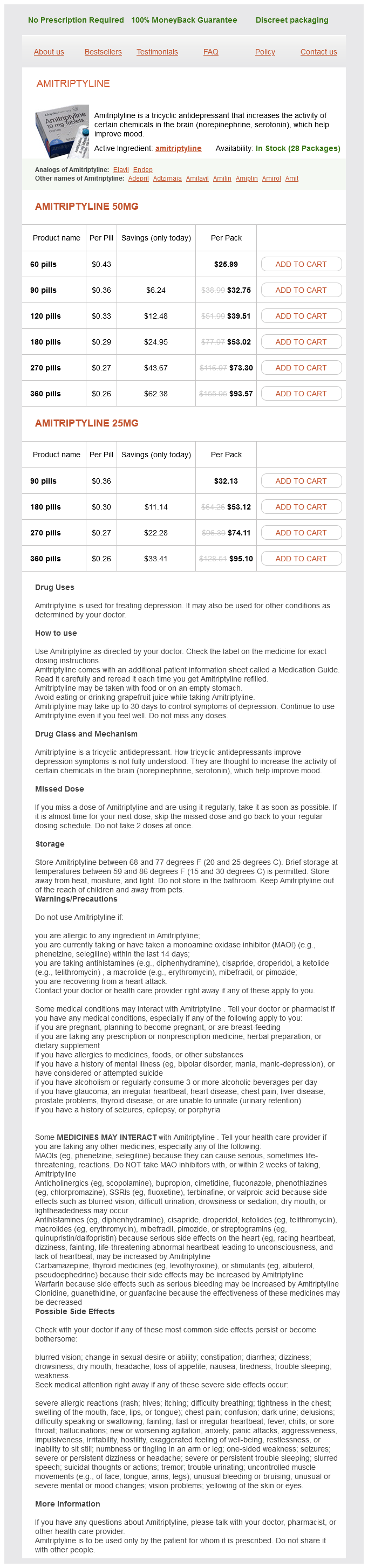
Amitriptyline Dosage and Price
Amitriptyline 50mg
- 60 pills - $25.99
- 90 pills - $32.75
- 120 pills - $39.51
- 180 pills - $53.02
- 270 pills - $73.30
- 360 pills - $93.57
Amitriptyline 25mg
- 90 pills - $32.13
- 180 pills - $53.12
- 270 pills - $74.11
- 360 pills - $95.10
Thus anxiety 24 hour hotline amitriptyline 25 mg online, division of the renal vein must be proximal to the entry of these vessels, and the vein cannot be divided after renal mobilization. In 95% of cases, the opening of numerous collateral channels allows the kidney to survive. The gonadal vein provides the principal route, anastomosing with the internal iliac veins, especially in women, but it is functional only if the valves are incompetent. The left adrenal vein by its connection with the inferior phrenic vein may bypass the renal vein. The renoazygolumbar channel is a very large collateral that could possibly handle the entire flow. This channel runs from the inferior or posterior border of the left renal vein across the left border of the aorta to divide into an inferior branch that drains into a lumbar vein and a superior branch that ascends to join a tributary of the hemiazygos vein. This channel joins the intravertebral plexuses that surround the nerve roots, and its failure accounts for the quadriplegia associated with stenosis of the left renal vein. In addition, the network in the perirenal tissue provides some collateral circulation by enlarging and draining into the inferior phrenic, subcostal, adrenal, lumbar, and gonadal veins as well as into the vena cava itself. Conversely, obstruction of the portal vein may be compensated for by flow through the left renal vein. Intrarenal Veins, Right Kidney the peritubular capillary plexus of veins about the collecting tubules in the medulla drains through the venae rectae into the arcuate veins. Small venules join the interlobular vein that courses with the corresponding interlobular artery. Stellate veins on the surface of the kidney communicate with the capsular venous drainage and so form a connection between the intrarenal and extrarenal drainage systems. They may be found along the grooves on the surface of the kidney; they represent the margins of fetal lobulation. The stellate veins and veins from the cortex drain into the interlobular veins, then into the arcuate veins that accompany the arcuate arteries. The interlobar veins join together to form two or three trunks that empty into the renal vein. The arcuate veins form interconnecting arches around the bases of the pyramids at the corticomedullary line. From the arches, large interlobar veins run centrally beside the pyramids to reach the distal portion of the calyceal necks, where they form another set of anastomotic collars around them. The posterior lobar veins draining the posterior portion of the kidney pass between the calyceal necks of the minor calyces to join an anterior lobar vein. Two sets of anterior and posterior lobar veins are usually found; one drains the superior portion, and the other drains the inferior portion of the kidney. The lobar veins unite within the renal sinus or at the lip of the hilum to form two trunks that pass anterior to the pelvis. The intrarenal veins, unlike the arteries, do not have a segmental arrangement, and in addition, they freely anastomose within the renal substance. Thus, they are not terminal but form longitudinal arcades at three levels: (1) between the stellate veins, (2) between the arcuate veins, and (3) between the interlobar veins. The anastomotic arrangement of the sets of veins permits ligation of major channels without fear of venous obstruction. The intrarenal veins are large; intraoperative section or injury during percutaneous procedures may result in considerable blood loss. Because the venous anastomoses that form collars about the necks of the calyces and the lobar veins curving around the calyces are much larger in the anterior portion of the kidney, the posterior approach that is taken to avoid the arteries is equally useful in avoiding the larger veins. Incisions into the wall of the calyx for plastic revision or for stone removal may inadvertently enter the veins crossing between the calyceal necks. This approach not only reduces arterial injury but also avoids entering the venous collar. For endoscopic incision of the ureteropelvic junction, it is important to appreciate that in two-fifths of cases an inferior tributary to the renal vein courses on the anterior aspect of the renal pelvis, making a lateral incision necessary. A retropelvic vein is present in two-thirds of cases, draining some of the posterior portion of the kidney. This 5-mm vein can be of surgical importance because it lies in the margin of the hilum, where it is susceptible to division during calicotomy and may be injured along with the posterior segmental artery during direct puncture of the renal pelvis. Capsular Veins the capsular veins communicate with small veins in the perirenal tissue that constitute a network of accessory veins in the perirenal fat. The accessory veins receive blood from adjacent muscles and from the adrenal gland and the diaphragm separately from retroperitoneal channels and drain it into the left renal vein. The superficial system lies immediately beneath the overlying perirenal fascia and peritoneum and drains into the deep system. The deep system, in turn, passes blood to the major veins that drain the renal parenchyma. Of the principal veins, the superior capsular veins exit from the perirenal space between the kidney and adrenal to drain into the adrenal vein. The inferior capsular veins come from the lower pole to drain into the gonadal vein or into a branch of the renal vein. With obstruction of the left renal vein, all these channels enlarge to form a dense network about the kidney that drains into the inferior phrenic, subcostal, adrenal, lumbar, and gonadal veins and into the inferior vena cava itself. Lymphatic vessels run from the plexuses into a dense basal network over the base of the pyramids, where the channels from the cortex join with those from the medulla to reach the region of the calyceal fornix. From there, the lymphatics run with the blood vessels around the calyceal necks to the renal sinus, where they empty into several large valved collectors lying on the surface of the pelvis and accompany the renal vein out of the hilum to terminate in a few nodes along the renal vessels and in the aortic nodes.
© 2025 Adrive Pharma, All Rights Reserved..