General Information about Aleve
One of the primary advantages of Aleve is its long-lasting relief. While other ache relievers may only last for a couple of hours, Aleve can provide relief for up to 12 hours, making it a popular choice for these with continual pain. This signifies that individuals can take it less regularly, which can assist scale back the danger of potential unwanted side effects associated with overuse of ache medicine.
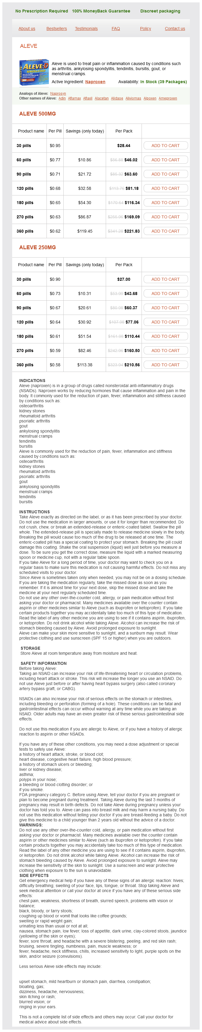
Aleve, additionally known by its generic name naproxen, is a non-steroidal anti-inflammatory drug (NSAID) commonly used to deal with quite lots of circumstances together with arthritis, ankylosing spondylitis, tendinitis, bursitis, gout, and menstrual cramps. It is on the market in each prescription and over-the-counter forms, making it easily accessible to those in need of fast-acting pain relief.
The energetic ingredient in Aleve, naproxen, works by blocking the production of certain chemical substances in the body that cause irritation and pain. This helps to reduce swelling and ease discomfort associated with various situations. Aleve is on the market in different types corresponding to tablets, liquid gels, and caplets, making it convenient for folks to choose the shape that works finest for them.
Pain is a universal expertise that we've all dealt with in some unspecified time in the future in our lives. From headaches to muscle cramps, ache can be a debilitating condition that affects our day by day actions and high quality of life. As a result, many individuals turn to over-the-counter medications to search out reduction. One such medicine is Aleve, a popular pain reliever that has been trusted by hundreds of thousands of people worldwide.
Another advantage of Aleve is that it comes in different strengths, which permits for personalised dosing based mostly on a person's wants. This means that for minor aches and pains, a decrease strength may be adequate, while a higher energy could additionally be needed for more severe circumstances. However, it is necessary to consult with a healthcare professional when figuring out the suitable dose as taking too much of any medication can have antagonistic results.
While Aleve is mostly protected and well-tolerated, it is important to focus on potential unwanted aspect effects. These may include stomach upset, heartburn, headache, nausea, and dizziness. In rare cases, it might also trigger severe side effects, together with liver or kidney issues, allergic reactions, and increased threat of heart attack or stroke. It is crucial to observe the recommended dosage and speak to a healthcare skilled if any regarding unwanted aspect effects occur.
It is a broadly known proven truth that managing pain is crucial in selling general well-being and Aleve performs a major role on this. Whether it's for acute or continual ache, Aleve has been confirmed to be an effective and safe option when taken as directed. In truth, a examine printed in the British Journal of Clinical Pharmacology found that Aleve was equally effective as other generally used ache relievers, but with a lower threat of side effects. This offers customers peace of thoughts figuring out they will find aid with out having to worry about potential adverse reactions.
In conclusion, Aleve is a trusted and effective ache reliever that has been offering reduction to hundreds of thousands of people for many years. Its long-lasting results and varied strengths make it a convenient possibility for those looking for focused reduction. However, as with all treatment, it's essential to make use of it responsibly and consult with a healthcare professional when wanted. With correct use, Aleve might help us handle pain and improve our quality of life. So the subsequent time you experience ache, consider Aleve as a secure and effective choice for reduction.
Molecular mechanisms of optic vesicle development: complexities pain management during shingles purchase aleve 250 mg without a prescription, ambiguities and controversies. Development and organization of the retina: cellular, molecular and functional perspectives, Semin Cell Dev Biol. Anterior eye development and ocular mesenchyme: new insights from mouse models and human diseases. Lens induction in vertebrates: variations on a conserved theme of signaling events. Secreted inducers in vertebrate eye development: more functions for old morphogens. Experimental studies on the development of the eye in amphibia, I: on the origin of the lens. Semaphorin3A/neuropilin-1 signaling acts as a molecular switch regulating neural crest migration during cornea development. Roles of cell-extrinsic growth factors in vertebrate eye pattern formation and retinogenesis. Establishing the pre-placodal region and breaking it into placodes with discrete identities. Early development of the cranial sensory nervous system: from a common field to individual placodes. The preplacodal region: an ectodermal domain with multipotential progenitors that contribute to sense organs and cranial sensory ganglia. Induction and specification of the vertebrate ectodermal placodes: precursors of the cranial sensory organs. From shared lineage to distinct functions: the development of the inner ear and epibranchial placodes. Molecular and tissue interactions governing induction of cranial ectodermal placodes. A better understanding of the development of the stomodeum and the breakdown of the oropharyngeal membrane has come from recent experiments on Xenopus embryos. Activation of shh and the inhibition of Wnt activity are required for the specification of the stomodeum and for regulation of its size. In contrast to the tympanic membrane, the oropharyngeal membrane does not contain a layer of mesenchyme between the ectodermal and endodermal epithelia. One of the first steps in breakdown of the oropharyngeal membrane is dissolution of the basement membrane, which requires shh activity. After dissolution of the basement membrane, the cells of the ectodermal and endodermal layers intercalate, resulting in a one-cell thick oropharyngeal membrane, which soon thereafter breaks down and opens a direct connection between the primitive gut and the outside. In the rostral midline is the frontonasal prominence, which is populated by mesenchymal cells derived from forebrain and some midbrain neural crest. On either side of the frontonasal prominence, ectodermal nasal placodes, which arose alongside the anterior neural ridge (see p. Farther caudally, the stomodeum is bounded by maxillary and mandibular processes, which are also filled with neural crestderived mesenchyme. The future cervical region is dominated by the pharyngeal apparatus, consisting of a series of pharyngeal pouches, arches, and clefts. Many components of the face, ears, and glands of the head and neck arise from the pharyngeal region. While vertebrates became more complex, the contributions of the neural crest to the head became much more prominent, and the face and many dermal (intramembranously formed) bones of the skull (dermocranium) were added. With the early evolution of the face, the most anterior of the branchial arches underwent a transformation to form the upper and lower jaws and two of the middle ear bones, the malleus and the incus. From structural and molecular aspects, the rostral (anteriormost) part of the head shows distinctly different characteristics from the pharyngeal region, as follows: 1. Formation of the forebrain and associated structures of the rostral part of the head depends on the actions of specific genes. Much of the connective tissue and skeleton of the rostral (phylogenetically newer) part of the head is derived from the neural crest. The anterior end of the notochord, terminating at the hypophysis, constitutes the boundary between the mesodermally derived chondrocranium and the more rostral neural crestderived chondrocranium. Neural crest cells are also prominent contributors to the ventral part of the pharyngeal region. The first part of this chapter provides an integrated view of early craniofacial development to show how the major components are interrelated. The remainder of the chapter concentrates on the development of the face, pharynx, and pharyngeal arch system. The chain of events between segmental patterns of gene expression and the appearance of morphological segmentation in parts of the cranial region is the subject of much current research. Cephalization begins with the rapid expansion of the rostral end of the neural plate. Very early, the future brain is the dominant component of the craniofacial region. Alternating with the pharyngeal grooves and pouches are paired masses of mesenchyme called pharyngeal (branchial) arches. The mesenchyme of the incipient musculature originates from mesoderm, specifically the somitomeres. Much of the remaining pharyngeal arch mesenchyme, especially that of the ventral part, is derived from the neural crest, whereas mesoderm makes various contributions to the dorsal pharyngeal arch mesenchyme.
Other reagents and solvents pain treatment guidelines pdf order aleve american express, procured from local sources, were of analytical grade. Antiulcer Activity and Experimental Design the assay was carried out with the methodology described by Almasaudi et al. All treatments were administrated by intragastric gavage for eight consecutive days, with the gastric ulcer induced on the last day with 80% ethanol solution (1 mL/animal). Group I (vehicle control) received the vehicle only (10 mL/kg body weight (bw) of 1% Tween-80 aqueous solution). For all other groups, an ulcer was induced on the last day of treatment, one hour after administering the corresponding compound. After 10 min, the stomachs were opened over the greater curvature and rinsed with saline solution (0. The gastric damage area (mm2) was determined with "Image J" image processing software. Stomach Tissue Preparation In a second experiment, another series of four groups of rats were formed. After the eight days of the corresponding treatments, the ulcer was induced and the rats were sacrificed (see previous section). The supernatants were divided into aliquots and conserved at -20 C until the biochemical analysis. The decrease in absorbance was measured after 1 and 2 min at 340 nm, with enzyme activity being directly proportional to the rate of change. The protein concentration in supernatants was established by the Bradford method [41], using bovine serum albumin as a standard. This assay involves the binding of Coomassie Brilliant Blue G-250 dye to proteins at r. When the dye binds to the protein, it is converted from an unstable form (red in color) to a stable unprotonated form (turning blue). After incubation for 15 min in boiling water, the samples were cooled and centrifuged at 1000 rpm for 10 min at 4 C. Subsequently, they were dehydrated by immersing them in ascending concentrations of alcohol solutions (70100%) and in paraffin. Slides of stomach slices of 45 m thickness were prepared and stained with hematoxylin and eosin (H&E) and then analyzed under light microscope at 20× and 40× for pathological changes, including necrosis, edema, vasocongestion, eosinophilic infiltration, and glandular damage. Statistical Analysis Statistical analysis was carried out with SigmaPlot version 12. Nine different proteins were identified that belong to phycobilisomes, which is a light-harvesting macromolecular complex (Table 2). The spot number (band of gel), accession number, protein name, unused, % coverture (% Cov), and molecular weight are reported. Other proteins were also detected (their specific data are summarized in Table S1). Effect of ExPhy on Ethanol-Induced Gastric Lesions the gastroprotective effect of pretreatment with ExPhy on ethanol-induced gastric lesions was determined (Table 3). The decrease in the ulcer index was also expressed as a percentage of protection, being 35. Pretreatment with omeprazole decreased the gastric lesions compared to the ulcer control. Pretreatment with ExPhy resulted in gastric lesions, characterized by focal areas of disruption in one-third of the mucosa, without a mucus layer in this zone. Effect of ExPhy and omeprazole on ulcer parameters in rats with ethanol-induced ulcers. H&E staining of rat gastric mucosa in ethanol-induced gastric ulcers (magnification at 20× and 40×). Discussion Considering the frequency of gastric ulcers in humans and the side effects and cost of some available synthetic drugs, the use of natural products represents an important alternative for many [43,44]. In this sense, Spirulina maxima and ExPhy have proven to be advantageous in the treatment of various ailments in lab animals and patients. Moreover, their absence of toxicity has been demonstrated by short- and long-term studies [45]. A determination was made of the effects of ExPhy in relation to some antioxidant and oxidative markers, along with protection against histopathological damage. Phycobilisomes are supramolecular complexes on the stromal surface of the thylakoid membrane in cyanobacteria. These complexes, which participate in trapping light energy and transferring it within the cell, can make up to 60% of the total protein [46,47]. The antioxidant potential reported for Am may be attributed to this major class of proteins. Phycobilisomes are constituted mainly by individual protein components denominated phycobiliproteins and linker polypeptides [48]. Phycobiliproteins consist of two different polypeptides (the and chains) that are covalently linked to bilin chromophores [49]. Interestingly, the analysis by mass spectrometry showed the presence of other cellular proteins (see the Supplementary Materials) that probably influenced the purity of phycobiliproteins in a minor way. Phycobiliproteins have attracted attention due to their special structure and potential therapeutic properties, either in a pure state or in protein extract. Excessive ethanol consumption is considered one of the risk factors for gastric ulcers in humans [61,62]. Its use in experimental animals allows for the evaluation of cytoprotective activity of potentially active products [63]. Different mechanisms of gastric cytoprotection have been suggested, including increased gastric mucosal blood flow, free radical scavenging, and stimulation of cell growth and repair [64]. In the current study, consistent with previous findings, administration of 80% ethanol solution by intragastric gavage produced marked damage in the gastric mucosa of rats, characterized mainly by elongated macroscopic lesions with intense hemorrhaging and hyperemia, as well as loss of mucus [35,65,66].
Aleve Dosage and Price
Aleve 500mg
- 30 pills - $28.44
- 60 pills - $46.02
- 90 pills - $63.60
- 120 pills - $81.18
- 180 pills - $116.34
- 270 pills - $169.09
- 360 pills - $221.83
Aleve 250mg
- 30 pills - $27.00
- 60 pills - $43.68
- 90 pills - $60.37
- 120 pills - $77.06
- 180 pills - $110.44
- 270 pills - $160.50
- 360 pills - $210.56
Getting axons onto the right path: the role of transcription factors in axon guidance laser pain treatment utah buy aleve 500 mg mastercard. Pattern formation in the vertebrate neural tube: a sonic hedgehog morphogen-regulated transcriptional network. Parasympathetic neurons originate from nerve-associated peripheral glial precursors. Siah regulation of Pard3A controls neuronal cell adhesion during germinal zone exit. Morphogen to mitogen: the multiple roles of hedgehog signaling in vertebrate neural development. Slit-mediated repulsion is a key regulator of motor axon pathfinding in the hindbrain. Mechanisms regulating the development of the corpus callosum and its agenesis in mouse and human. The initial appearance of the cranial nerves and related neuronal migration in staged human embryos. The timing and sequence of appearance of neuromeres and their derivatives in staged human embryos. Hoxa2- and rhombomere-dependent development of the mouse facial somatosensory map. Otx dose-dependent integrated control of anteroposterior and dorso-ventral patterning of midbrain. Oligodendrocyte precursors migrate along vasculature in the developing nervous system. Wnt won the war: antagonistic role of Wnt over shh controls dorso-ventral patterning of the vertebrate neural tube. Integrity of developing spinal motor columns is regulated by neural crest derivatives at motor exit points. How does Fgf signaling from the isthmic organizer induce midbrain and cerebellum development Sonic hedgehog functions through dynamic changes in temporal competence in the developing forebrain. Among the new transcription factors upregulated in specified neural crest precursor cells are snail-1,-2 (formerly called slug), Twist, and Foxd-3, which are instrumental in allowing the neural crest cells to undergo an epitheliomesenchymal transformation. These cells then break free from the neural epithelium and then migrate away as mesenchymal cells. Neural crest cells break free from the neural tube in the trunk at the level of the last-formed somite or the neural plate in the head by changing their shape and properties from those of typical neuroepithelial cells to those of mesenchymal cells. These molecules remain downregulated during migration, but after neural crest cells have completed their migrations and have differentiated into certain structures. In the head, where closure of the neural plate has not yet occurred, neural crest cells must penetrate the basal lamina underlying the neural plate. This is accomplished by the production of enzymes that degrade components of the basal lamina and by sending out processes that penetrate the basal lamina. In the trunk, neural crest cells do not leave the neuroepithelium until after the neural tube has formed. They do not, however, have to contend with penetrating a basal lamina because the dorsal part of the neural tube does not form a basal lamina until after emigration of the crest cells. The neural crest, the existence of which has been recognized for more than a century, forms an exceptionally wide range of cell types and structures, including several types of nerves and glia, connective tissue, bones, and pigment cells. Its importance and prominence are such that the neural crest has often been called the fourth germ layer of the body. Not until adequate methods of marking neural crest cells became available-first with isotopic labels and subsequently with stable biological markers, monoclonal antibodies, intracellular dyes, and genetic markers-did the neural crest become one of the most widely studied components of the vertebrate embryo. More recently, emphasis has shifted to studies on the mouse, especially for dissecting molecular controls, but it appears that most of the information on the biology of the neural crest derived from birds can be applied to mammalian embryos. Some important syndromes and malformations are based on abnormalities of the neural crest. Tracing the history of the neural crest in any region involves consideration of the following: (1) its origin, induction, and specification; (2) epithelial-tomesenchymal transformation and emigration from the neural tube; (3) migration; and (4) differentiation. Each of these phases in the development of the generic neural crest is covered before neural crest development in specific regions of the body is considered. In addition to neural crest precursors, the neural plate border contains several types of progenitor cells, such as progenitors of the ectodermal placodes in the anterior region. In response to these inductive signals, cells at the border of the neural plate activate genes coding for several transcription factors, including Msx-1,-2, Dlx-5, and Pax-3/Pax-7. They also activate another set of genes (Foxd-3, Sox-10, and Ets-1), which specify the neural crest progenitor cells within the neural plate border. In this environment, the cells undergo extensive migrations along several well-defined pathways. These migrations are determined by intrinsic properties of the neural crest cells and features of the external environment encountered by the migrating cells. One of its main functions is to prevent premature differentiation of the migrating cells. Neural crest migration is influenced by a variety of molecules residing in the extracellular matrix. Although the presence of a basal lamina can inhibit their emigration from the neural tube, neural crest cells often prefer to migrate along basal laminae, such as those of the surface ectoderm or neural tube, after they have left the neural tube. Components of the extracellular * Snail-2 is also expressed during gastrulation by cells of the epiblast after they have entered the walls of the primitive streak and are about to leave as mesenchymal cells of the mesoderm germ layer.
© 2025 Adrive Pharma, All Rights Reserved..